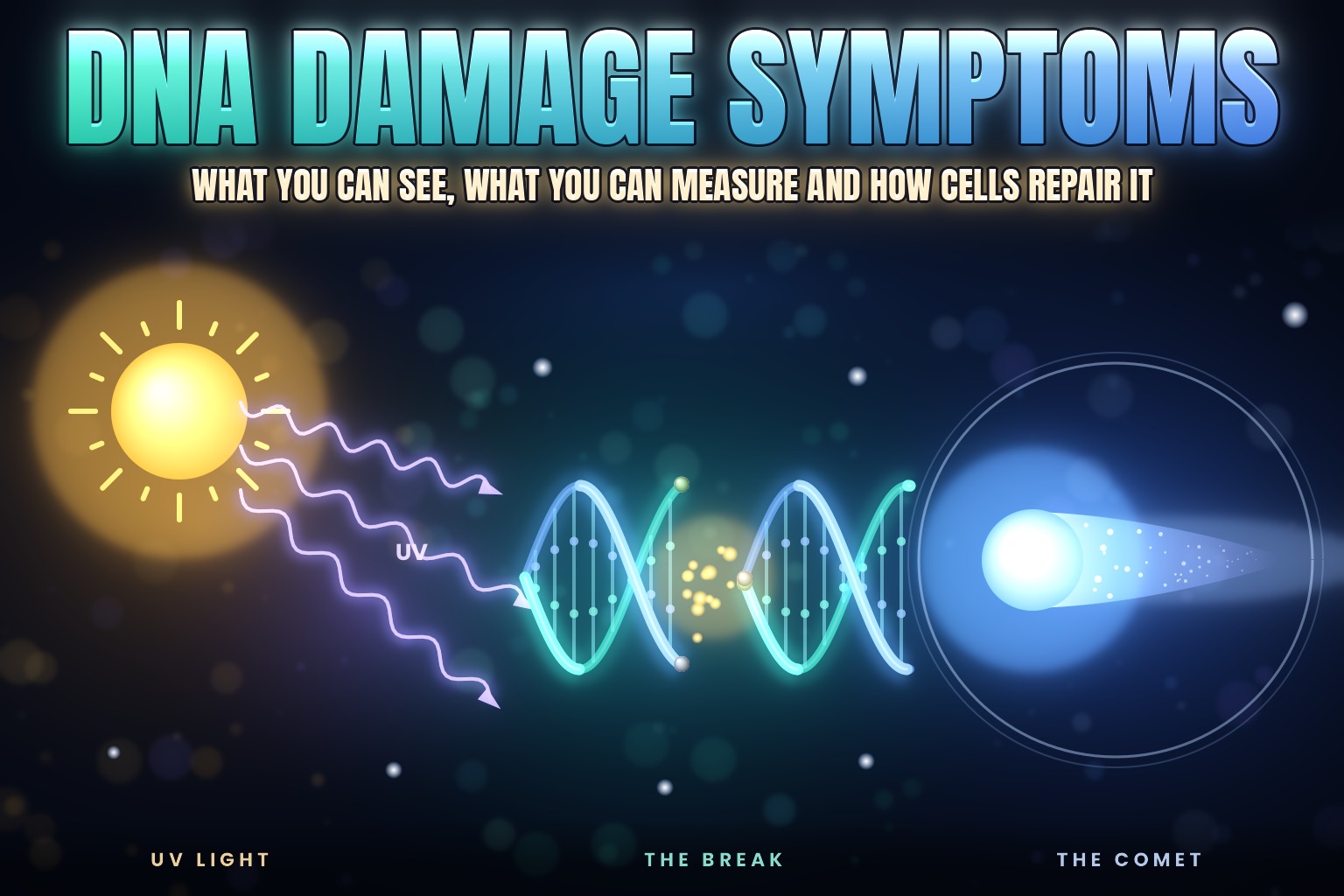
DNA falls apart on its own. It decays so steadily that it sets a limit on how far back scientists can recover DNA from fossils.
The same chemistry runs in your cells all the time, and repair systems work against it. DNA damage is something you see and measure rather than feel, so here’s the sign you can see, the tests that measure the rest, what raises and lowers the markers, and what changes with age.
What DNA Damage Is
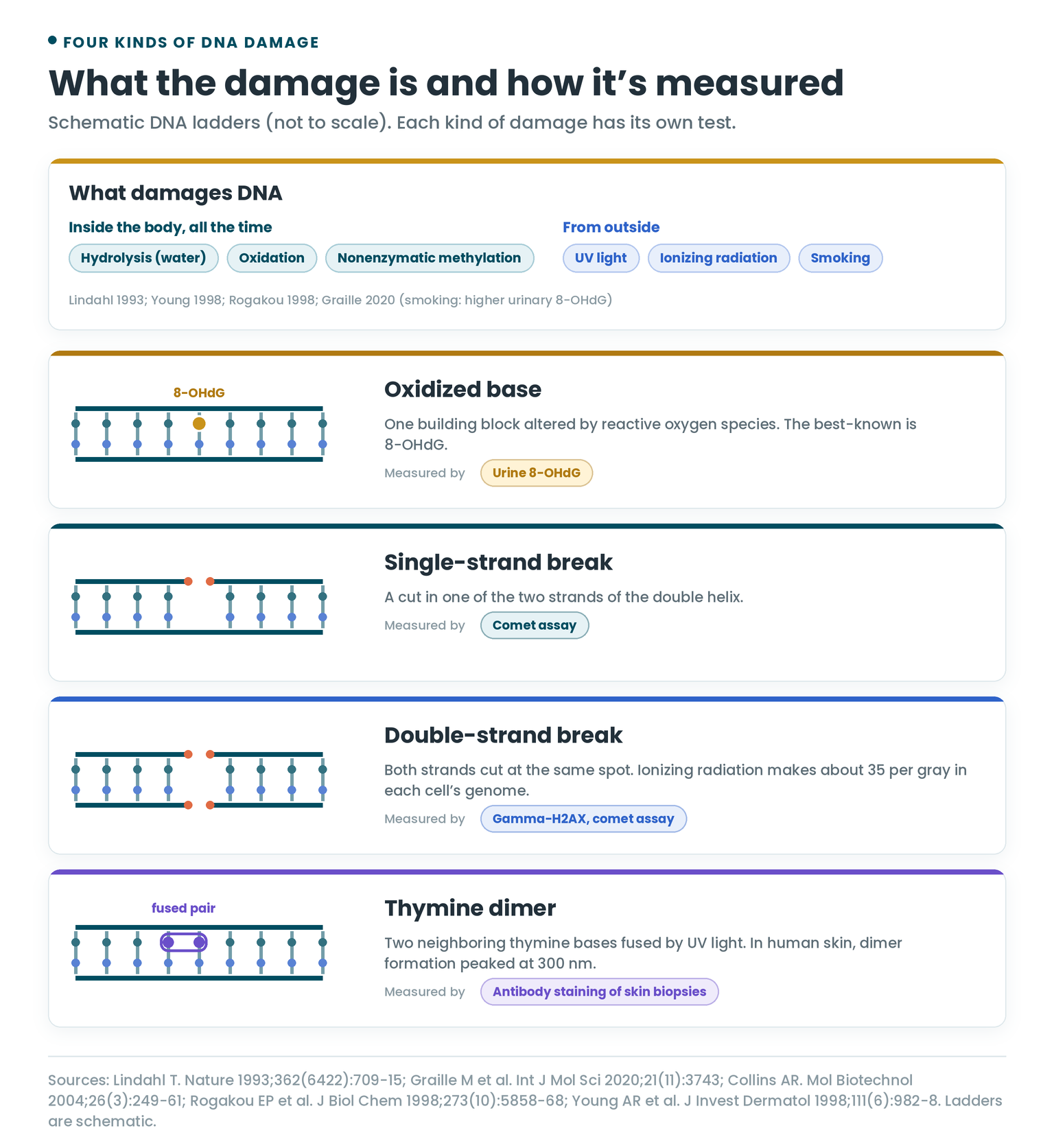
DNA carries your genetic information, but chemically it’s a fragile molecule. Three reactions chip away at it at significant rates inside the body:
- Hydrolysis: water breaking bonds in the DNA.
- Oxidation: reactive oxygen species altering its building blocks.
- Nonenzymatic methylation: small methyl groups attaching where they shouldn’t.
Spontaneous DNA decay is thought to be a major factor in mutations and aging.
Oxidative Stress
Free radicals are released as a by-product of respiration, the way cells burn fuel with oxygen, and the body’s antioxidant system mops them up. When reactive oxygen species build up faster than they’re cleared, that imbalance is called oxidative stress. In excess, they oxidize proteins, fats and DNA.
That makes some oxidative damage part of normal life. A 1993 study used an enzyme that cuts DNA at oxidized bases, combined with the comet assay (more on that below), and found significant oxidative damage in freshly drawn white blood cells from normal, healthy people.
Common Types of DNA Damage
| Type | What it is | Common cause | How it’s measured |
|---|---|---|---|
| Oxidized base | One of DNA’s four building blocks altered by oxidation. The best-known is 8-hydroxy-2-deoxyguanosine (8-OHdG). | Reactive oxygen species | Urine 8-OHdG; comet assay with a repair enzyme |
| Single-strand break | A cut in one of the two strands of the double helix | Various, including hard exercise and sleep loss | Comet assay |
| Double-strand break | Both strands cut at the same spot | Ionizing radiation, including CT scans | Gamma-H2AX, comet assay |
| Thymine dimer | Two neighboring thymine bases fused together | Ultraviolet (UV) light, both UVB and UVA | Antibody staining of skin biopsies; mass spectrometry |
| 6-4 photoproduct | A second kind of UV lesion linking two neighboring bases | UV light | Antibody staining of skin biopsies |
How Cells Repair DNA Damage
Specific repair processes counteract hydrolysis, oxidation and methylation, and most DNA damage is repaired. Two traces of that repair work turn out to be useful:
- Oxidized products leave in urine. That’s what makes a urine test for DNA damage possible.
- PARP enzymes burn NAD+. PARPs help repair DNA, and they use up NAD+ as they work. That ties DNA damage to NAD+ levels.
Repair isn’t the same in everyone. How fast and how completely cells repair comes up again and again below: after sun exposure, after a CT scan, and with age.
The Sign You Can See: Sunburn
Sunburn redness is a sign of DNA damage you can see with your own eyes.
A 1998 study irradiated previously unexposed buttock skin in volunteers with UV light at seven wavelengths, from 280 to 360 nm, at 0.5 to 3 minimal erythema doses. One minimal erythema dose is the smallest dose that turns the skin red. Biopsies taken right after exposure were stained for thymine dimers in four regions of the epidermis, from the basal layer up.
- Peak damage at 300 nm. All four dimer action spectra peaked at 300 nm.
- Deep reach. At 300 nm, dimers turned up even in the dermis, below the epidermis. Solar UVB (about 295 to 320 nm) was more damaging to the basal layer, where new skin cells are made, than predicted from measurements on skin samples outside the body.
- Redness tracks the damage. The wavelengths that made dimers lined up with the wavelengths that made the same volunteers’ skin red. The authors concluded that DNA is a major chromophore for sunburn redness between 280 and 340 nm, meaning it’s a main molecule absorbing the UV that sets it off.
How Fast Skin Repairs Sun Damage
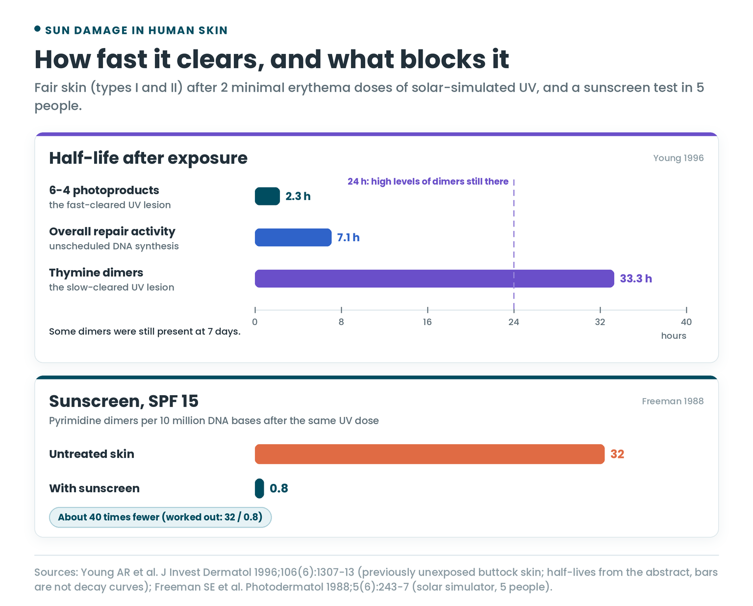
A 1996 study from the same group timed the repair in people with fair skin (skin types I and II). Previously unexposed buttock skin got 2 minimal erythema doses of solar-simulated UV, and biopsies tracked the damage over the following days:
- Thymine dimers cleared slowly. Their half-life was 33.3 hours. High levels were still there at 24 hours, and some were still present at 7 days.
- 6-4 photoproducts cleared fast, with a half-life of 2.3 hours.
- Overall repair activity in the skin, measured as unscheduled DNA synthesis, fell with a half-life of 7.1 hours. It tracked the 6-4 photoproducts more closely than the dimers.
The timing of the sunburn lined up with the fast repair. The onset of redness was inversely related to 6-4 photoproduct repair, and the authors suggested that this repair process is what sets the redness off.
UVA Damages DNA Too
UVB (290 to 320 nm) is the part of sunlight DNA absorbs directly. UVA (320 to 400 nm) is more abundant and isn’t directly absorbed by DNA, but it still does damage. A 2006 study measured DNA damage in human skin exposed to UVB or UVA, using HPLC with tandem mass spectrometry:
- UVA made dimers too. Cyclobutane pyrimidine dimers, the typical UVB lesion, formed in significant amounts under UVA, by a different mechanism.
- More dimers than oxidation. Under UVA, these dimers outnumbered 8-oxo-dG (the same lesion as 8-OHdG), the most common oxidative lesion in skin.
- Slower to clear. Dimers made by UVA were removed more slowly than those made by UVB.
- Weak natural shield. Compared with skin cells grown from the same donors, whole skin protected its cells well against UVB damage but only weakly against UVA.
For what peptides have shown in skin research, see Peptides in Skincare.
How DNA Damage Is Measured
Beyond sunburn, DNA damage is tracked in the lab. Three tests do most of the work.
In Urine: 8-OHdG
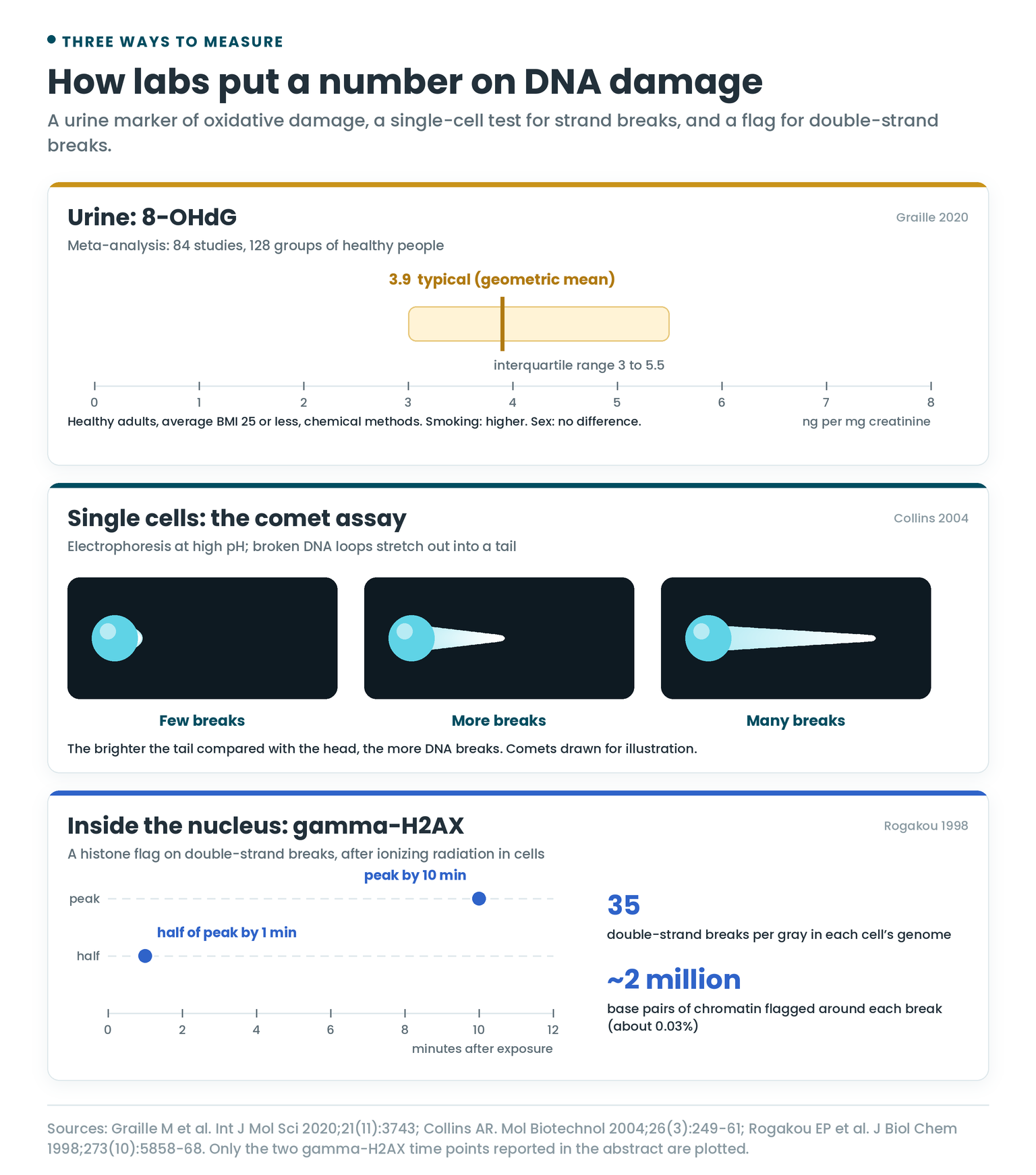
Because oxidized products of repair are excreted in urine, urinary 8-OHdG is used as a marker of oxidative DNA damage.
A 2020 meta-analysis set out to define what’s normal. It screened 1,246 articles, kept 84, and pooled 128 groups of healthy people:
- Typical level: 3.9 ng per mg of creatinine (geometric mean, interquartile range 3 to 5.5) in healthy adults with an average BMI of 25 or less, measured by chemical methods.
- Smoking: linked to significantly higher urinary 8-OHdG by chemical methods.
- Sex: no difference between men and women.
Results are given per mg of creatinine to correct for how concentrated the urine is.
In Single Cells: The Comet Assay
The comet assay, also called single-cell gel electrophoresis, measures DNA strand breaks in individual cells:
- Cells are set in a thin gel on a microscope slide and burst open with detergent and salt, leaving the DNA behind as tightly coiled loops.
- An electric current runs through the slide at high pH. The likely reason the next step works: loops with a break uncoil and are free to stretch toward the positive pole.
- Under a fluorescence microscope, each cell looks like a comet. The brighter the tail compared with the head, the more breaks.
Adding repair enzymes that cut DNA at specific kinds of damage makes the test more sensitive and more specific, because each lesion they find becomes an extra break in the tail. Measuring how much damage remains at intervals after exposure shows repair as it happens.
What’s Normal on the Comet Assay
The hCOMET project set out to answer that. It pooled comet results from 19,320 people in 105 studies, run by 44 labs in 26 countries between 1999 and 2019:
- Reference ranges: it reported interquartile ranges for the standard comet readouts, including tail intensity, tail length and tail moment.
- Age: a small effect in some datasets, with more DNA damage in the oldest age groups.
- Sex and smoking: no effect on comet results, though the authors noted the data held few heavy smokers.
- Exposures: highly significant differences for most of the exposures studied, which is why the authors called the comet assay an excellent biomarker of exposure.
Score a Comet
Try reading comets yourself. Estimate how much of each cell’s DNA is in the tail, then reveal the answer. A second panel shows how a repair time course reads.
Inside the Nucleus: Gamma-H2AX
When both strands of DNA break, the cell flags the spot. A histone protein called H2AX, part of the spool that DNA wraps around, gets a phosphate group added at one specific site. The marked form is called gamma-H2AX.
The 1998 study that identified it found:
- It’s fast. After ionizing radiation in cell cultures, gamma-H2AX reached half its peak within 1 minute and its peak within 10 minutes.
- It’s big. Each double-strand break involved about 2 million base pairs’ worth of chromatin, roughly 0.03% of the genome.
The authors called gamma-H2AX formation a rapid and sensitive cellular response to double-strand breaks.
Gamma-H2AX in People: CT Scans
A 2005 study counted gamma-H2AX foci, the stained spots that mark breaks, in white blood cells from people having CT scans of the chest or abdomen:
- Breaks tracked the dose. The number of double-strand breaks rose in a straight line with the scan’s dose-length product, a measure of both the dose and how much of the body was scanned.
- Repair back to baseline. Sampled for up to a day afterward, healthy people repaired the breaks back to background levels.
- One outlier. A patient who had previously had severe side effects from radiotherapy carried several times more foci at each time point, and tests on their cells grown in the lab (fibroblasts) confirmed a substantial repair defect.
What Raises DNA Damage Markers
UV light, ionizing radiation and smoking all show up in the tests above. Two everyday factors have human data too.
Sleep Loss and Night Shifts
A 2019 study of 49 healthy full-time doctors took blood after three nights of adequate sleep, and again the morning after an overnight on-site shift for the 24 who worked them:
- At baseline, doctors who worked overnight shifts already had more DNA breaks and lower activity of DNA repair genes than those who didn’t, with large effect sizes (Cohen’s d of 1.48 and 1.47).
- After a night without sleep, their DNA breaks went up further (d = 0.87) and repair gene activity went down (d = 0.90).
It was an observational study, so it shows an association between disrupted sleep and DNA damage.
Exhaustive Exercise
In a 1995 study, people ran on a treadmill until exhaustion, and their white blood cells were tested with the comet assay before and 24 hours after:
- Every runner showed a clear increase in DNA strand breaks at 24 hours.
- Short-term vitamins (multivitamins, or vitamin E at 3 doses of 800 mg) gave a significantly smaller increase in some of them.
- Vitamin E at 1,200 mg a day for 14 days before the run clearly reduced the damage in all of them and prevented it entirely in 4 of 5.
Malondialdehyde, a marker of fat oxidation, fell significantly after the 14 days of vitamin E. The authors concluded the breaks come from oxidative stress after exhaustive exercise.
DNA Damage and Aging
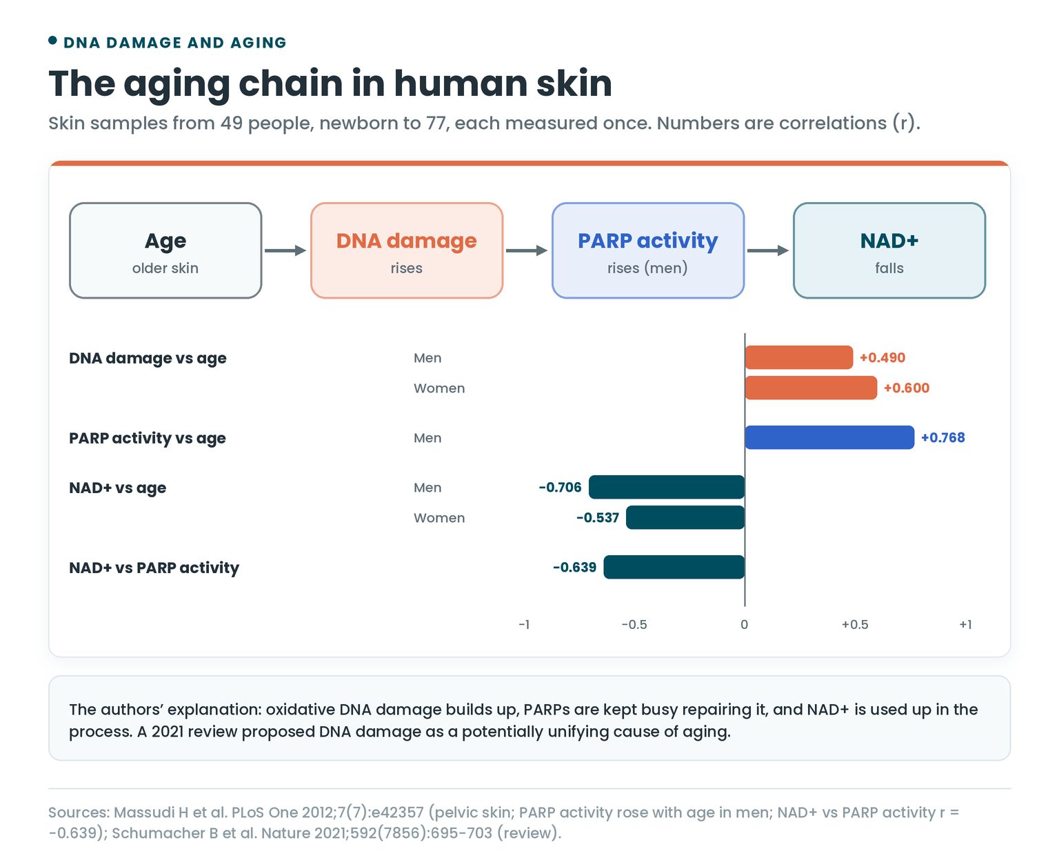
In skin samples from 49 people, aged from newborn to 77, DNA damage rose with age in both men (r = 0.490) and women (r = 0.600). Over the same span, NAD+ fell. In men, PARP activity climbed with age (r = 0.768), and higher PARP activity went with lower NAD+ (r = −0.639).
The authors’ explanation: oxidative DNA damage accumulates, PARPs are kept busy repairing it, and NAD+ gets drained in the process. Those are correlations from people measured once, at different ages.
A 2021 review in Nature took the idea further. Its authors pulled together evidence that DNA damage affects most, if not all, features of aging, and proposed it as a potentially unifying cause of aging.
For how NAD+ is measured, see NAD Test. For the mouse research on NAD+ and DNA repair, see David Sinclair’s NMN.
Repair Slows With Age
Three studies of human cells point the same way:
- UV repair capacity. In skin cells grown from donors aged from their first to their tenth decade, the ability to repair UV-damaged DNA fell 0.6% per year. In blood cell lines from the same age range, UV-induced mutations rose 0.6% per year. Over 50 years, that adds up to roughly 30% (my arithmetic, assuming a straight line).
- Break processing. Gamma-H2AX foci in connective-tissue cells (fibroblasts) and white blood cells became more common with age, and repair proteins arrived at breaks more slowly in cells from older donors.
- Blood-forming stem cells. In healthy people of different ages, unrepaired double-strand breaks in blood-forming stem and progenitor cells rose with age, along with shorter telomeres, though most breaks weren’t at the telomeres. Variation between people was largest in middle age, and each person’s repair capacity appeared to set how much damage built up. Healthy people over 70 showed only a modest rise and kept their repair efficiency, which led the authors to suggest that a healthy lifestyle may slow the natural aging process.
What Lowers DNA Damage Markers
Three lines of human evidence point to less damage or better protection: one in living skin, one in blood cells, and one in skin cells and skin samples in the lab.
Sunscreen
In a 1988 study, the skin of 5 people was exposed to UV from a solar simulator, with and without a chemical sunscreen rated SPF 15. Biopsies were then tested for pyrimidine dimers, the family that thymine dimers belong to:
- Untreated skin: 32 dimers per 10 million DNA bases.
- Sunscreen-treated skin, same UV dose: 0.8 per 10 million bases.
That’s about 40 times fewer (my arithmetic).
Fruit and Antioxidants
A 2001 trial gave volunteers a single 0.5-liter drink of kiwifruit juice, with water as the control, then tested their white blood cells (lymphocytes) with the comet assay:
- More resistant DNA. After the kiwifruit drink, the cells’ DNA held up better against oxidative damage from hydrogen peroxide than after water.
- Same baseline. The cells’ existing level of DNA damage stayed the same.
- Beyond vitamin C. In the test tube, a kiwifruit extract protected DNA better than vitamin C at the same concentration, though neither protected at the highest concentrations tested. The authors concluded the fruit’s antioxidant effect wasn’t entirely down to its vitamin C.
Nicotinamide (Vitamin B3) and Repair
Nicotinamide is a form of vitamin B3 and a precursor of NAD+, which cells need to make energy (ATP). Since DNA repair runs on energy, a 2013 lab study tested whether nicotinamide would speed it up after low-dose solar-simulated UV:
- More repair, faster. In human skin cells (keratinocytes), nicotinamide increased both the share of cells carrying out excision repair and the repair rate in each cell.
- Fewer lesions. In those cells and in human skin samples, nicotinamide reduced both UV dimers and 8-oxo-dG, and the reduction appeared to come from faster repair.
That makes it two different repair pathways boosted at once. For the NAD+ precursors studied in people, see Best NAD+ Supplements.
Doses Used in the Research
- UV light on human skin: 0.5, 1, 2 and 3 minimal erythema doses at 280, 290, 300, 310, 320, 340 and 360 nm (Young 1998).
- Solar-simulated UV on fair skin: 2 minimal erythema doses (Young 1996).
- Ionizing radiation in cells: about 1% of H2AX marked per gray, from about 35 double-strand breaks per gray in each cell’s genome (Rogakou 1998).
- Sunscreen: a chemical sunscreen rated SPF 15 against an equal UV dose from a solar simulator (Freeman 1988).
- Kiwifruit juice: a single 0.5-liter drink, with water as the control (Collins 2001).
- Vitamin E before exhaustive exercise: 3 doses of 800 mg short-term, or 1,200 mg a day for 14 days (Hartmann 1995).
Frequently Asked Questions
What are the symptoms of DNA damage?
DNA damage is measured rather than felt. The visible sign is sunburn: the UV wavelengths that caused DNA damage in human skin were the same ones that turned it red.
What causes DNA damage?
Water, oxidation and stray methylation damage DNA all the time inside the body. UV light, ionizing radiation and smoking add more, and in human studies so did sleep loss and exhaustive exercise.
How do you test for DNA damage?
The three main tests are urinary 8-OHdG for oxidative damage, the comet assay for strand breaks in single cells, and gamma-H2AX for double-strand breaks.
What is a normal 8-OHdG level?
In a meta-analysis of healthy adults with an average BMI of 25 or less, urinary 8-OHdG averaged 3.9 ng per mg of creatinine (interquartile range 3 to 5.5) by chemical methods.
Can DNA damage be repaired?
Yes. Specific repair processes counteract the damage that happens all the time, and most of it is repaired. After CT scans, healthy people repaired double-strand breaks in their white blood cells back to background levels within a day.
How long does sun damage to DNA take to repair?
In fair human skin, thymine dimers from UV had a half-life of 33.3 hours, and some were still present after 7 days. The other main UV lesion, the 6-4 photoproduct, cleared with a half-life of 2.3 hours.
Does UVA damage DNA?
Yes. In human skin, UVA produced the same dimers as UVB, more of them than oxidative lesions, and they were removed more slowly than UVB dimers.
Does sunscreen prevent DNA damage?
In a study of 5 people, an SPF 15 sunscreen cut UV-induced pyrimidine dimers from 32 to 0.8 per 10 million DNA bases.
Does lack of sleep cause DNA damage?
In 49 doctors, a night of on-call sleep deprivation went with more DNA breaks and lower DNA repair gene activity the next morning.
Does exercise cause DNA damage?
Exhaustive treadmill running increased DNA strand breaks in white blood cells 24 hours later in every runner tested, and 14 days of vitamin E beforehand reduced it.
Does DNA damage increase with age?
In human skin, yes. DNA damage rose with age in both men and women, while NAD+ fell, and studies of human cells found repair slowing with age.
Stay sharp,
Joe Mars
The Peptide Report
P.S. Reading up on DNA repair, NAD+ or aging? Bring your questions to the Peptides & Biohacking Hub on Skool.
References
- Lindahl T. Instability and decay of the primary structure of DNA. Nature. 1993;362(6422):709-715. doi:10.1038/362709a0
- Collins AR, Duthie SJ, Dobson VL. Direct enzymic detection of endogenous oxidative base damage in human lymphocyte DNA. Carcinogenesis. 1993;14(9):1733-1735. doi:10.1093/carcin/14.9.1733
- Graille M, Wild P, Sauvain JJ, et al. Urinary 8-OHdG as a biomarker for oxidative stress: a systematic literature review and meta-analysis. Int J Mol Sci. 2020;21(11):3743. doi:10.3390/ijms21113743
- Young AR, Chadwick CA, Harrison GI, et al. The similarity of action spectra for thymine dimers in human epidermis and erythema suggests that DNA is the chromophore for erythema. J Invest Dermatol. 1998;111(6):982-988. doi:10.1046/j.1523-1747.1998.00436.x
- Young AR, Chadwick CA, Harrison GI, et al. The in situ repair kinetics of epidermal thymine dimers and 6-4 photoproducts in human skin types I and II. J Invest Dermatol. 1996;106(6):1307-1313. doi:10.1111/1523-1747.ep12349031
- Mouret S, Baudouin C, Charveron M, et al. Cyclobutane pyrimidine dimers are predominant DNA lesions in whole human skin exposed to UVA radiation. Proc Natl Acad Sci U S A. 2006;103(37):13765-13770. doi:10.1073/pnas.0604213103
- Collins AR. The comet assay for DNA damage and repair: principles, applications, and limitations. Mol Biotechnol. 2004;26(3):249-261. doi:10.1385/MB:26:3:249
- Milić M, Ceppi M, Bruzzone M, et al. The hCOMET project: international database comparison of results with the comet assay in human biomonitoring. Baseline frequency of DNA damage and effect of main confounders. Mutat Res Rev Mutat Res. 2021;787:108371. doi:10.1016/j.mrrev.2021.108371
- Rogakou EP, Pilch DR, Orr AH, et al. DNA double-stranded breaks induce histone H2AX phosphorylation on serine 139. J Biol Chem. 1998;273(10):5858-5868. doi:10.1074/jbc.273.10.5858
- Löbrich M, Rief N, Kühne M, et al. In vivo formation and repair of DNA double-strand breaks after computed tomography examinations. Proc Natl Acad Sci U S A. 2005;102(25):8984-8989. doi:10.1073/pnas.0501895102
- Cheung V, Yuen VM, Wong GTC, Choi SW. The effect of sleep deprivation and disruption on DNA damage and health of doctors. Anaesthesia. 2019;74(4):434-440. doi:10.1111/anae.14533
- Hartmann A, Niess AM, Grünert-Fuchs M, et al. Vitamin E prevents exercise-induced DNA damage. Mutat Res. 1995;346(4):195-202. doi:10.1016/0165-7992(95)90035-7
- Massudi H, Grant R, Braidy N, et al. Age-associated changes in oxidative stress and NAD+ metabolism in human tissue. PLoS One. 2012;7(7):e42357. doi:10.1371/journal.pone.0042357
- Schumacher B, Pothof J, Vijg J, Hoeijmakers JHJ. The central role of DNA damage in the ageing process. Nature. 2021;592(7856):695-703. doi:10.1038/s41586-021-03307-7
- Moriwaki S, Ray S, Tarone RE, et al. The effect of donor age on the processing of UV-damaged DNA by cultured human cells: reduced DNA repair capacity and increased DNA mutability. Mutat Res. 1996;364(2):117-123. doi:10.1016/0921-8777(96)00029-8
- Sedelnikova OA, Horikawa I, Redon C, et al. Delayed kinetics of DNA double-strand break processing in normal and pathological aging. Aging Cell. 2008;7(1):89-100. doi:10.1111/j.1474-9726.2007.00354.x
- Rübe CE, Fricke A, Widmann TA, et al. Accumulation of DNA damage in hematopoietic stem and progenitor cells during human aging. PLoS One. 2011;6(3):e17487. doi:10.1371/journal.pone.0017487
- Freeman SE, Ley RD, Ley KD. Sunscreen protection against UV-induced pyrimidine dimers in DNA of human skin in situ. Photodermatol. 1988;5(6):243-247. PubMed 2854891
- Collins BH, Horská A, Hotten PM, et al. Kiwifruit protects against oxidative DNA damage in human cells and in vitro. Nutr Cancer. 2001;39(1):148-153. doi:10.1207/S15327914nc391_20
- Surjana D, Halliday GM, Damian DL. Nicotinamide enhances repair of ultraviolet radiation-induced DNA damage in human keratinocytes and ex vivo skin. Carcinogenesis. 2013;34(5):1144-1149. doi:10.1093/carcin/bgt017