
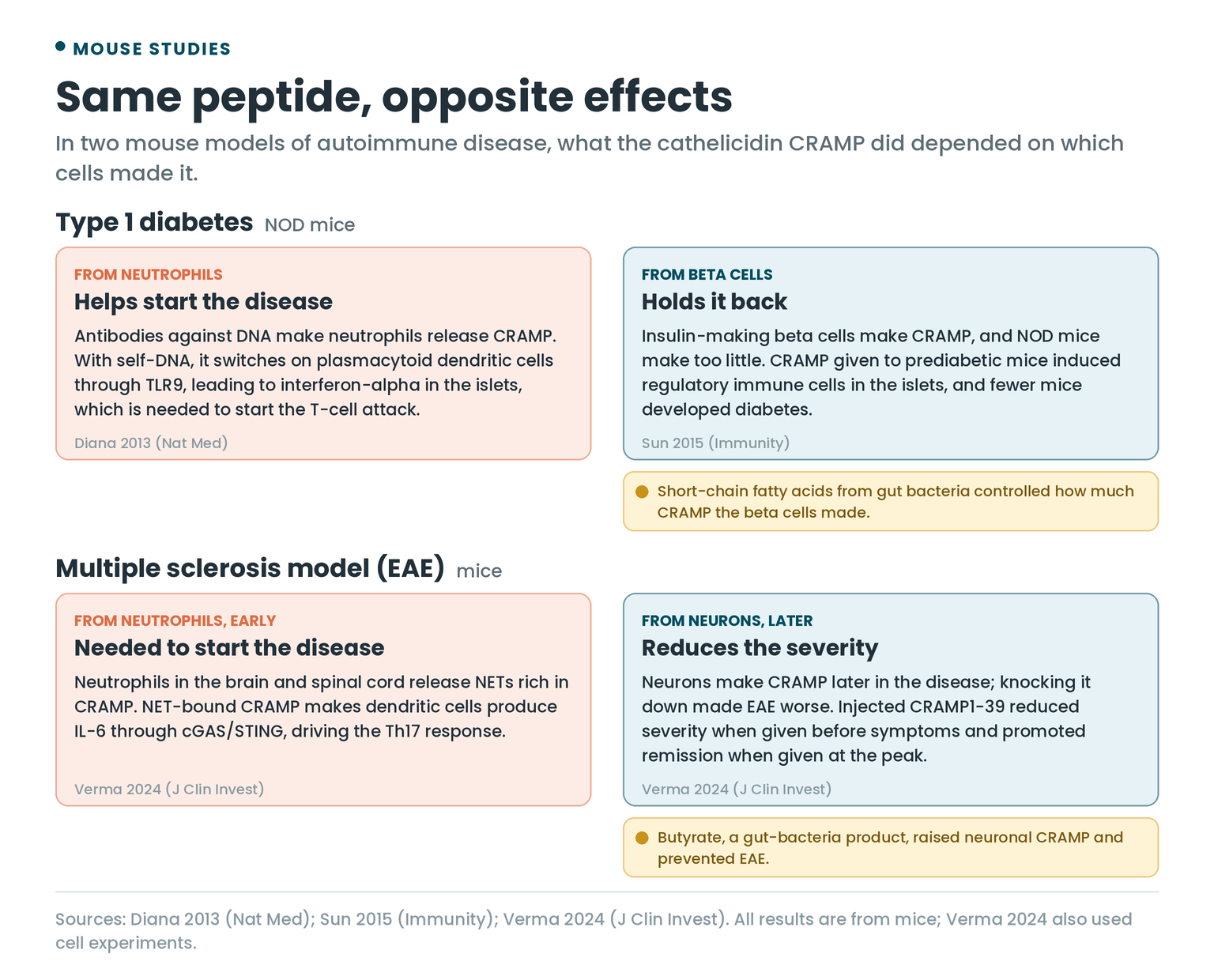
In 2013, a team including Julien Diana showed that a peptide called CRAMP is part of the spark that starts type 1 diabetes in mice. Two years later, Diana’s own lab gave CRAMP to the same strain of mice, and fewer of them developed diabetes.
Both papers are right. In the first, neutrophils released the peptide. In the second, it came from the insulin-making cells themselves, or from an injection. Where an immune peptide comes from, and what it’s carrying when it arrives, decides what it does to your immune system. That’s the thread through everything below.
What Immune Peptides Are
When people talk about peptides and the immune system, the ones your body makes for itself are mostly antimicrobial peptides, also called host defense peptides. They’re short and positively charged, and they fight infection two ways: by attacking microbes directly and by changing how your own immune cells respond. A 2020 review in Nature Reviews Drug Discovery covered both jobs. The second one is what this article is about.
Humans make two main families:
- Cathelicidin. You have exactly one. Its gene, described in 1996, is the only member of the cathelin gene family in the human genome. Neutrophils and epithelial cells make it as a larger protein, hCAP18. In neutrophils, an enzyme called proteinase 3 cuts the active 37-amino-acid peptide, LL-37, off the end once the protein has been released from the cell. Mice have their own version, CRAMP, which is why that name runs through the mouse studies.
- Defensins. The alpha-defensins include neutrophil defensin 1, also called alpha-defensin 1. The beta-defensins are made by the linings of the skin, the salivary glands and the airways. I went through the airway ones in Peptides and Lung Health.
How Host Defense Peptides Steer Immune Cells
Killing microbes is the job these peptides are named for. Their immune work falls into four kinds.
Calling Immune Cells In
A 1999 paper in Science showed that human beta-defensins attract immature dendritic cells and memory T cells. They do it through CCR6, a chemokine receptor those cells carry, which gives the defensins a way to pull the adaptive immune system toward an infection. A year later, the same research group showed that LL-37 attracts neutrophils, monocytes and T cells through a different receptor, FPRL1.
Turning Inflammation Down
In 2006, Neeloffer Mookherjee and colleagues tested LL-37 on human immune cells stimulated with LPS, the endotoxin in the outer membrane of Gram-negative bacteria. At 1 µg/mL or less, a range the authors call physiological, LL-37 blocked the release of TNF-alpha from monocytic cells. It cut the move of NF-kB, which switches on many inflammatory genes, into the cell nucleus by half or more. In blood immune cells it almost completely stopped the cytokine burst triggered by LPS and by other Toll-like receptor signals.
It was selective. LL-37 left alone the genes that wind inflammation back down, and it didn’t block the response to TNF-alpha or IL-1beta themselves.
The same lab then built a peptide around the idea. IDR-1, an “innate defense regulator”, doesn’t kill bacteria at all. Given to mice anywhere from 48 hours before to 6 hours after infection, it still protected them against MRSA, vancomycin-resistant Enterococcus and Salmonella, by raising the chemokines that recruit monocytes while lowering inflammatory cytokines. The protection disappeared when monocytes and macrophages were removed.
Shaping T Cells
With TGF-beta1 present, cathelicidin pushes T cells away from the Th1 type and into Th17, the IL-17-making subset, and it protects Th17 cells, but not Th1 cells, from dying. Mice without cathelicidin made weaker Th17 responses during inflammation, though not at rest. The 2021 study that found this also showed neutrophils releasing cathelicidin inside mouse lymph nodes, where T cells are trained.
Carrying Your Own DNA and RNA Into Dendritic Cells
This is where autoimmunity comes in.
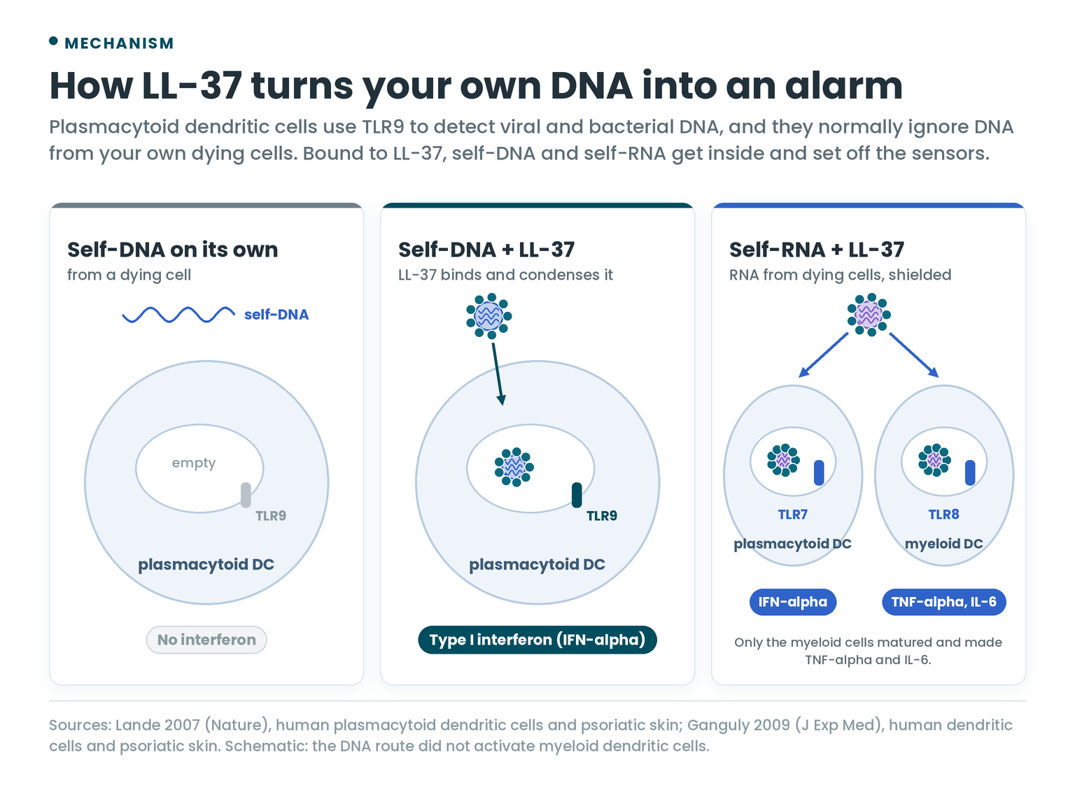
Plasmacytoid dendritic cells are built to spot viral and bacterial DNA and answer with type I interferon. Normally they ignore DNA from your own dying cells. In 2007, Roberto Lande, Michel Gilliet and colleagues found that LL-37 breaks that rule. It binds self-DNA into condensed clumps that get delivered into the compartments inside the cell where the DNA sensor TLR9 sits, and TLR9 fires. They identified LL-37 as the key factor switching these cells on in psoriasis.
Two years later the same group showed LL-37 does the same with RNA from dying cells. It shields the RNA from being broken down outside the cell and carries it in, where it triggers TLR7 in plasmacytoid dendritic cells and TLR8 in classical (myeloid) dendritic cells. The RNA route also made the myeloid cells mature and release TNF-alpha and IL-6, which the DNA route didn’t.
Antimicrobial Peptides in Autoimmune Diseases
Here’s how that plays out disease by disease. The human evidence comes mostly from patients’ skin, blood and joints. The cause-and-effect experiments are mostly in mice and rats.
Psoriasis
Normal skin holds almost no LL-37 or beta-defensin 2. In a 2002 New England Journal of Medicine study, both were abundant in the top layer of the epidermis in every psoriasis patient tested, and significantly lower in the lesions of atopic dermatitis. The two peptides together killed Staphylococcus aureus, and the authors suggested the shortfall may explain why eczema skin gets infected with it so easily.
Genetics points the same way. Edward Hollox and colleagues counted copies of the beta-defensin genes on chromosome 8 in two cohorts: 179 Dutch people with psoriasis against 272 controls, and 319 German people with psoriasis against 305 controls. In both, carrying more copies went with a higher risk of psoriasis.
Then in 2014, Lande’s group found that LL-37 is also a target. Two-thirds of patients with moderate-to-severe plaque psoriasis carried T cells that recognize LL-37. Those T cells made interferon-gamma, the CD4 ones also made Th17 cytokines, they were found in lesional skin, and their numbers in the blood tracked disease activity. So in psoriasis LL-37 works at two levels: it switches on dendritic cells, and T cells recognize it as a target.
Systemic Lupus Erythematosus
Lupus is marked by chronic activation of plasmacytoid dendritic cells and by autoantibodies against the cell nucleus. Two papers in the same 2011 issue of Science Translational Medicine tied both to neutrophils.
The link is the NET, short for neutrophil extracellular trap: a web of DNA that a neutrophil throws out, studded with its antimicrobial peptides. Lande and colleagues found complexes of neutrophil antimicrobial peptides and self-DNA in lupus patients’ blood, made in the form of NETs, and they switched on plasmacytoid dendritic cells through TLR9. The patients made autoantibodies against both the DNA and the peptides, and lupus neutrophils released more NETs than healthy ones, more still when those antibodies were added. The second paper, in children with lupus, found NETs carrying large amounts of LL-37 and HMGB1 that drove high interferon-alpha output, depending on both the DNA and TLR9.
Two more pieces followed. In 2013, NETs and LL-37 were shown to switch on the inflammasome in macrophages, releasing IL-1beta and IL-18, more strongly in macrophages from lupus patients. IL-18 in turn made neutrophils release more NETs, a loop that feeds itself.
In a 2020 study, 45% of the lupus patients had T cells that responded strongly to LL-37, tracking their anti-LL-37 antibodies and disease activity. Unlike the Th17-type cells in psoriasis, these looked like T follicular helper cells, the type that helps B cells make antibodies, and in the lab they did push B cells to make anti-LL-37 antibodies. They reacted most to citrullinated LL-37, a form in which enzymes have converted the amino acid arginine to citrulline, and the researchers found plenty of it in lupus skin and kidney.
Rheumatoid Arthritis
Cathelicidins are strongly raised in the joint lining of people with rheumatoid arthritis compared with healthy joints, mostly in neutrophils and in macrophages and osteoclasts. In rats with pristane-induced arthritis, the rat cathelicidin rose at the same time as cell death, interferon-alpha and autoantibodies. The 2013 authors concluded the peptides might help start the disease or keep it going.
The same NET machinery shows up here. Neutrophils from the blood and joint fluid of people with rheumatoid arthritis form more NETs than those from healthy people or people with osteoarthritis. The NETs push citrullinated proteins out of the cell, the targets of the anti-citrullinated protein antibodies (ACPAs) considered a key step in rheumatoid arthritis, and NET formation tracked ACPA levels. IL-17A and TNF-alpha triggered NETs, and the NETs made synovial fibroblasts more inflammatory.
A piece of LL-37 pulled the other way. IG-19, an internal segment of LL-37, lowered disease severity in mice with collagen-induced arthritis, cut their antibodies against type II collagen, reduced the immune cells in their joints, prevented cartilage breakdown and lowered inflammatory cytokines. IDR-1018, a peptide derived from a cow cathelicidin, didn’t do the same in the same model.
Type 1 Diabetes
The two studies from the top of this article come from NOD mice, a strain bred to develop autoimmune diabetes.
In the 2013 study, normal beta-cell death in young female NOD mice drew B-1a cells, neutrophils and plasmacytoid dendritic cells into the pancreas. The B-1a cells made antibodies against DNA, and those antibodies made neutrophils release CRAMP, which bound self-DNA. The DNA, the antibodies and CRAMP together switched on the dendritic cells through TLR9, and the dendritic cells made interferon-alpha in the islets. Removing the B-1a cells, the neutrophils or the interferon-making dendritic cells blocked the start of the T-cell attack and the diabetes.
In the 2015 study, the beta cells themselves made CRAMP, and NOD mice made too little of it. CRAMP given to prediabetic NOD mice induced regulatory immune cells in the islets. Beta-cell CRAMP depended on short-chain fatty acids made by gut bacteria, and changing the mice’s gut microbiota changed their CRAMP and their islet inflammation.
Multiple Sclerosis
The mouse model for multiple sclerosis is experimental autoimmune encephalomyelitis (EAE), and Th17 cells have been widely implicated in both. In a 2022 study, neutrophils, microglia and blood vessel cells released cathelicidin throughout EAE. It boosted Th17 cells in the lymph nodes and their switch to interferon-gamma production in the spinal cord, and mice lacking cathelicidin were protected from severe disease. The same cell types made cathelicidin in active brain lesions from people with MS.
A 2024 study from Diana’s lab found both directions inside one disease:
- Early. Neutrophils recruited into the brain and spinal cord made NETs rich in CRAMP, and those NETs were needed to start EAE. NET-bound CRAMP made dendritic cells produce IL-6 through the cGAS/STING pathway, driving the Th17 response.
- Later. Neurons made CRAMP that reduced the severity. Knocking it down in neurons made the disease worse. A recombinant version, CRAMP1-39, injected before symptoms reduced severity and injected at the peak promoted remission, and injected into the spinal fluid space it lowered a blood marker of nerve damage.
- Gut link. Butyrate, a gut-bacteria product, raised neuronal CRAMP through the FFAR2 and FFAR3 receptors and prevented EAE.
Sjögren’s Syndrome
Sjögren’s goes the other way from psoriasis. A 2009 study expected more beta-defensin in the inflamed salivary glands and found less: beta-defensins 1 and 2 were lower than in normal glands, and beta-defensin 2 dropped further.
The alpha-defensins show up instead. In a 2009 protein survey of minor salivary glands from 6 patients and 6 matched controls, alpha-defensin 1 was one of six proteins found only in the patients’ glands. A 2021 study looked at the antibody-antigen complexes in saliva and found neutrophil defensin 1 in them for 6 of 9 Sjögren’s patients, among the antigens specific to Sjögren’s when compared with 7 people with dry mouth (xerostomia). With myeloperoxidase, elastase and cathepsin G also on the list, all neutrophil proteins, the authors suggested repeated neutrophil destruction may be part of the disease.
LL-37 in People: A Leg Ulcer Trial
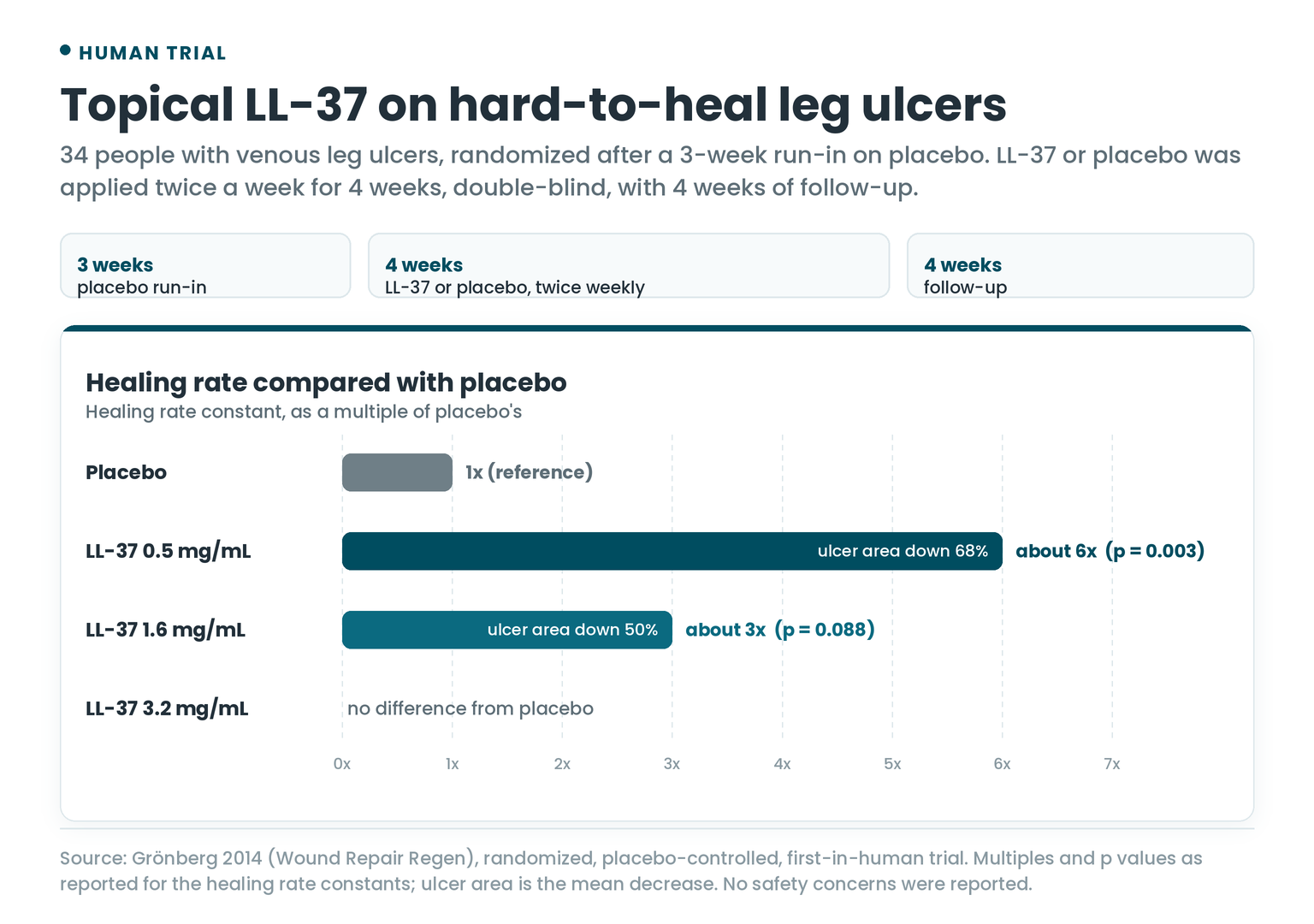
LL-37 was first given to people in a 2014 randomized, placebo-controlled trial on hard-to-heal venous leg ulcers. It enrolled 34 people. After a 3-week run-in on placebo, they got LL-37 at 0.5, 1.6 or 3.2 mg/mL, or placebo, applied twice a week for 4 weeks under double-blind conditions, then 4 weeks of follow-up.
- 0.5 mg/mL: a healing rate about six times placebo’s (p = 0.003), and the average ulcer area fell 68%.
- 1.6 mg/mL: about three times placebo’s (p = 0.088, short of statistical significance), with a 50% fall in ulcer area.
- 3.2 mg/mL: no difference from placebo.
There were no safety concerns, local or systemic.
For an immune peptide tested in vaccine, sepsis and COVID-19 trials, I went through thymosin alpha 1 in Thymosin Alpha 1 and Bodybuilding.
The Findings by Disease
The disease findings from this article, sorted by what each one showed the peptide doing.
The peptide ledger
Pick a disease. Each finding sits in the lane for what it showed: the peptide driving the disease, holding it back, or turning up in patients. Tap a card for the details.
Badges show where each finding comes from: people (tissue, blood or cells from patients), animals, or lab cells.
Doses Used in the Research
- LL-37 on venous leg ulcers (people): 0.5, 1.6 or 3.2 mg/mL applied twice a week for 4 weeks.
- LL-37 on human immune cells (lab): 1 µg/mL or less was enough to dampen the LPS response.
- IDR-1 (mice): given from 48 hours before to 6 hours after infection, locally or into the bloodstream.
- CRAMP1-39 (EAE mice): injected into the abdomen on day 7 or day 15 after the disease was induced, or into the spinal fluid space on day 9.
Frequently Asked Questions
How do peptides modulate the immune system?
Host defense peptides like LL-37 and the defensins call immune cells in, turn some inflammatory responses down, steer T cells toward particular types and carry DNA and RNA into dendritic cells. Which of those happens depends on which cells made the peptide and what it’s bound to.
Does LL-37 boost or calm the immune system?
Both, depending on the setting. In lab studies it dampened the cytokine response to bacterial endotoxin, while in psoriasis and lupus it turns self-DNA and self-RNA into triggers for interferon.
What is the link between LL-37 and psoriasis?
LL-37 is abundant in psoriatic skin, where it carries self-DNA and self-RNA into dendritic cells. Two-thirds of people with moderate-to-severe plaque psoriasis also have T cells that recognize LL-37.
What are NETs?
Neutrophil extracellular traps are webs of DNA that neutrophils release, studded with antimicrobial peptides like LL-37. In lupus they switch on interferon-making dendritic cells and become targets for autoantibodies.
Can antimicrobial peptides protect against autoimmune disease?
In mice, some have. CRAMP lowered diabetes in NOD mice and promoted remission in EAE, and IG-19, a piece of LL-37, calmed collagen-induced arthritis.
Has LL-37 been tested in people?
Yes, on hard-to-heal venous leg ulcers. In a 2014 randomized trial of 34 people, 0.5 mg/mL healed about six times faster than placebo, 1.6 mg/mL about three times faster but short of statistical significance, and 3.2 mg/mL no differently, with no safety concerns.
Stay sharp,
Joe Mars
The Peptide Report
P.S. Reading up on LL-37 or the defensins? Bring your questions to the Peptides & Biohacking Hub on Skool and we’ll go through the papers together.
References
- Diana J, Simoni Y, Furio L, et al. Crosstalk between neutrophils, B-1a cells and plasmacytoid dendritic cells initiates autoimmune diabetes. Nat Med. 2013;19(1):65-73. doi:10.1038/nm.3042
- Sun J, Furio L, Mecheri R, et al. Pancreatic β-cells limit autoimmune diabetes via an immunoregulatory antimicrobial peptide expressed under the influence of the gut microbiota. Immunity. 2015;43(2):304-317. doi:10.1016/j.immuni.2015.07.013
- Mookherjee N, Anderson MA, Haagsman HP, Davidson DJ. Antimicrobial host defence peptides: functions and clinical potential. Nat Rev Drug Discov. 2020;19(5):311-332. doi:10.1038/s41573-019-0058-8
- Gudmundsson GH, Agerberth B, Odeberg J, et al. The human gene FALL39 and processing of the cathelin precursor to the antibacterial peptide LL-37 in granulocytes. Eur J Biochem. 1996;238(2):325-332. doi:10.1111/j.1432-1033.1996.0325z.x
- Yang D, Chen Q, Schmidt AP, et al. LL-37, the neutrophil granule- and epithelial cell-derived cathelicidin, utilizes formyl peptide receptor-like 1 (FPRL1) as a receptor to chemoattract human peripheral blood neutrophils, monocytes, and T cells. J Exp Med. 2000;192(7):1069-1074. doi:10.1084/jem.192.7.1069
- Sørensen OE, Follin P, Johnsen AH, et al. Human cathelicidin, hCAP-18, is processed to the antimicrobial peptide LL-37 by extracellular cleavage with proteinase 3. Blood. 2001;97(12):3951-3959. doi:10.1182/blood.v97.12.3951
- Ong PY, Ohtake T, Brandt C, et al. Endogenous antimicrobial peptides and skin infections in atopic dermatitis. N Engl J Med. 2002;347(15):1151-1160. doi:10.1056/NEJMoa021481
- Kaneda Y, Yamaai T, Mizukawa N, et al. Localization of antimicrobial peptides human beta-defensins in minor salivary glands with Sjögren’s syndrome. Eur J Oral Sci. 2009;117(5):506-510. doi:10.1111/j.1600-0722.2009.00667.x
- Yang D, Chertov O, Bykovskaia SN, et al. Beta-defensins: linking innate and adaptive immunity through dendritic and T cell CCR6. Science. 1999;286(5439):525-528. doi:10.1126/science.286.5439.525
- Mookherjee N, Brown KL, Bowdish DM, et al. Modulation of the TLR-mediated inflammatory response by the endogenous human host defense peptide LL-37. J Immunol. 2006;176(4):2455-2464. doi:10.4049/jimmunol.176.4.2455
- Scott MG, Dullaghan E, Mookherjee N, et al. An anti-infective peptide that selectively modulates the innate immune response. Nat Biotechnol. 2007;25(4):465-472. doi:10.1038/nbt1288
- Minns D, Smith KJ, Alessandrini V, et al. The neutrophil antimicrobial peptide cathelicidin promotes Th17 differentiation. Nat Commun. 2021;12(1):1285. doi:10.1038/s41467-021-21533-5
- Lande R, Gregorio J, Facchinetti V, et al. Plasmacytoid dendritic cells sense self-DNA coupled with antimicrobial peptide. Nature. 2007;449(7162):564-569. doi:10.1038/nature06116
- Ganguly D, Chamilos G, Lande R, et al. Self-RNA-antimicrobial peptide complexes activate human dendritic cells through TLR7 and TLR8. J Exp Med. 2009;206(9):1983-1994. doi:10.1084/jem.20090480
- Hollox EJ, Huffmeier U, Zeeuwen PL, et al. Psoriasis is associated with increased beta-defensin genomic copy number. Nat Genet. 2008;40(1):23-25. doi:10.1038/ng.2007.48
- Lande R, Botti E, Jandus C, et al. The antimicrobial peptide LL37 is a T-cell autoantigen in psoriasis. Nat Commun. 2014;5:5621. doi:10.1038/ncomms6621
- Lande R, Ganguly D, Facchinetti V, et al. Neutrophils activate plasmacytoid dendritic cells by releasing self-DNA-peptide complexes in systemic lupus erythematosus. Sci Transl Med. 2011;3(73):73ra19. doi:10.1126/scitranslmed.3001180
- Garcia-Romo GS, Caielli S, Vega B, et al. Netting neutrophils are major inducers of type I IFN production in pediatric systemic lupus erythematosus. Sci Transl Med. 2011;3(73):73ra20. doi:10.1126/scitranslmed.3001201
- Kahlenberg JM, Carmona-Rivera C, Smith CK, Kaplan MJ. Neutrophil extracellular trap-associated protein activation of the NLRP3 inflammasome is enhanced in lupus macrophages. J Immunol. 2013;190(3):1217-1226. doi:10.4049/jimmunol.1202388
- Lande R, Palazzo R, Gestermann N, et al. Native/citrullinated LL37-specific T-cells help autoantibody production in systemic lupus erythematosus. Sci Rep. 2020;10(1):5851. doi:10.1038/s41598-020-62480-3
- Hoffmann MH, Bruns H, Bäckdahl L, et al. The cathelicidins LL-37 and rCRAMP are associated with pathogenic events of arthritis in humans and rats. Ann Rheum Dis. 2013;72(7):1239-1248. doi:10.1136/annrheumdis-2012-202218
- Khandpur R, Carmona-Rivera C, Vivekanandan-Giri A, et al. NETs are a source of citrullinated autoantigens and stimulate inflammatory responses in rheumatoid arthritis. Sci Transl Med. 2013;5(178):178ra40. doi:10.1126/scitranslmed.3005580
- Chow LN, Choi KY, Piyadasa H, et al. Human cathelicidin LL-37-derived peptide IG-19 confers protection in a murine model of collagen-induced arthritis. Mol Immunol. 2014;57(2):86-92. doi:10.1016/j.molimm.2013.08.011
- Smith KJ, Minns D, McHugh BJ, et al. The antimicrobial peptide cathelicidin drives development of experimental autoimmune encephalomyelitis in mice by affecting Th17 differentiation. PLoS Biol. 2022;20(8):e3001554. doi:10.1371/journal.pbio.3001554
- Verma SC, Enée E, Manasse K, et al. Cathelicidin antimicrobial peptide expression in neutrophils and neurons antagonistically modulates neuroinflammation. J Clin Invest. 2024;135(3):e184502. doi:10.1172/JCI184502
- Hjelmervik TO, Jonsson R, Bolstad AI. The minor salivary gland proteome in Sjögren’s syndrome. Oral Dis. 2009;15(5):342-353. doi:10.1111/j.1601-0825.2009.01531.x
- Yamane K, Nakamura H, Hamasaki M, et al. Immune complexome analysis reveals the presence of immune complexes and identifies disease-specific immune complex antigens in saliva samples from patients with Sjögren’s syndrome. Clin Exp Immunol. 2021;204(2):212-220. doi:10.1111/cei.13574
- Grönberg A, Mahlapuu M, Ståhle M, Whately-Smith C, Rollman O. Treatment with LL-37 is safe and effective in enhancing healing of hard-to-heal venous leg ulcers: a randomized, placebo-controlled clinical trial. Wound Repair Regen. 2014;22(5):613-621. doi:10.1111/wrr.12211