Your lungs hold about 480 million alveoli, and every breath reaches them through airways lined with peptides your own cells make. Some of those peptides kill bacteria. Some keep the airway surface wet. A copy of one surfactant protein was built to keep premature babies’ air sacs open.
That’s where the lung peptide research starts. From there it runs through smokers’ lungs, scarred lungs and the ICU. A few peptides made it into trials with hundreds of patients; others are still in mice. Here’s what each one did, layer by layer.
One Breath, Five Layers
Follow a breath in and it passes five places where peptides have been studied. Each section below takes one of them.
- The airway surface. A thin film of liquid coats your airways. The lining fills it with antimicrobial peptides and with SPLUNC1, a protein that keeps the film from drying out.
- The airway walls and their scaffolding. When smoke and enzymes break down collagen and elastin, the fragments become peptide signals that call in more inflammatory cells. This is where the COPD research sits.
- The air sacs. Surfactant keeps the alveoli open, and a synthetic peptide built to copy surfactant protein B was tested in two trials that enrolled more than 1,500 premature babies. When the alveoli flood, as in ARDS, a peptide that helps pump the fluid back out has been tested in ventilated patients.
- The tissue between the air sacs. In pulmonary fibrosis, scar builds up here. Caveolin-1 peptides, Ac-SDKP, thymosin beta-4 and GHK-Cu have all been tested against it in mice, and one caveolin-1 peptide has reached patients.
- The blood vessels. VIP relaxes lung arteries and calms lung immune cells, and BPC-157 has rat data on the pulmonary circulation.
The Peptides Your Airways Make Themselves
Defensins and cathelicidins are the main families of antimicrobial peptides in airway secretions, Pieter Hiemstra wrote in a 2007 review. They’re small, positively charged peptides made by the airway lining, and besides killing microbes directly they shape immune responses and wound repair.
Defensins: Antibiotics That Need the Right Salt
In 1997, Goldman and colleagues used grafts of human airway tissue to work out why cystic fibrosis airways can’t kill bacteria. The airway surface fluid from cystic fibrosis grafts carried abnormally high salt and failed to kill bacteria. The peptide behind the killing turned out to be human beta-defensin 1 (hBD-1), which the lining makes throughout the airways in both normal and cystic fibrosis lungs. Its activity against Pseudomonas aeruginosa depends on salt, so the high salt in cystic fibrosis airway fluid switches it off. Blocking it with antisense wiped out the bacteria-killing activity of normal airway fluid.
A year later the same lab found a second one, hBD-2, in the airway lining, the submucosal glands and the airway surface fluid. It kills a broad range of bacteria, it’s salt sensitive too, and it works together with lysozyme and lactoferrin.
SPLUNC1: Keeping the Airway Surface Wet
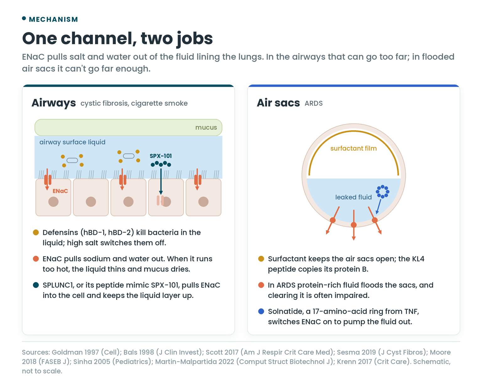
Mucus only moves if the airway surface stays hydrated. The epithelial sodium channel, ENaC, pulls sodium and water out of that layer. In cystic fibrosis it runs too hot, and the result is dried-out mucus, slower mucus clearance and repeated infections.
In healthy lungs a secreted protein called SPLUNC1 keeps ENaC in check. Cystic fibrosis sputum holds much less of it, and the proteases in that sputum break it down quickly. Cigarette smoke damages it as well: in a 2018 study, smoke-derived aldehydes modified SPLUNC1’s cysteines and made it clump irreversibly, so it could no longer pull ENaC off the cell surface.
SPX-101 is a peptide built to copy SPLUNC1’s ENaC-regulating action, without the cysteines. It binds ENaC, pulls all three of its subunits into the cell, and cuts the channel’s current. Inhaled once a day, it lifted survival in mice bred with an overactive ENaC to more than 90%, and it sped up mucus transport in those mice and in a sheep model of cystic fibrosis. It stayed intact and active after exposure to cystic fibrosis sputum, and after cigarette smoke it still restored airway surface hydration in cell cultures.
The same channel comes back in the ARDS section below, where the goal flips and you want ENaC working harder.
Peptides for Preterm Lungs: Copying Surfactant Protein B
Surfactant is the film that lowers surface tension inside the alveoli so they don’t collapse each time you breathe out. Babies born very early don’t make enough of it, which is respiratory distress syndrome (RDS). Lucinactant is a fully synthetic surfactant with no animal-derived components. Its active peptide, sinapultide, is 21 amino acids long, built from repeating leucine and lysine units (the KL4 peptide), and designed to mimic human surfactant protein B.
Two trials published together in 2005 tested it:
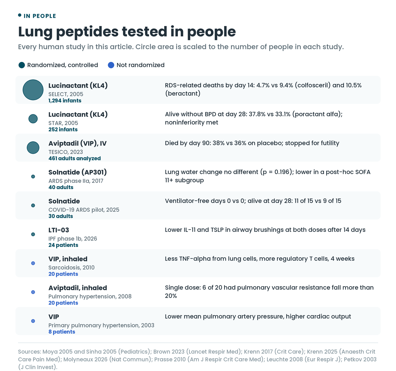
- SELECT (Fernando Moya and colleagues). 1,294 very preterm infants weighing 600 to 1,250 g got lucinactant, colfosceril (a phospholipid-only synthetic) or beractant (a bovine surfactant) within 20 to 30 minutes of birth. RDS at 24 hours was 39.1% on lucinactant against 47.2% on colfosceril, and not significantly different from beractant (33.3%). RDS-related deaths by day 14 were 4.7% on lucinactant against 9.4% on colfosceril and 10.5% on beractant.
- STAR (Sunil Sinha and colleagues). 252 infants born at 24 to 28 weeks got lucinactant or poractant alfa, a pig-derived surfactant. Survival without bronchopulmonary dysplasia at day 28 was 37.8% against 33.1%, which met the trial’s noninferiority margin. Deaths through day 28 were 11.8% against 16.1%.
In preterm lambs ventilated for four hours, lucinactant dampened lung and whole-body inflammation and preserved lung structure better than poractant alfa, beractant or no surfactant.
Peptides for Emphysema and COPD
PGP: When Broken Collagen Becomes a Signal
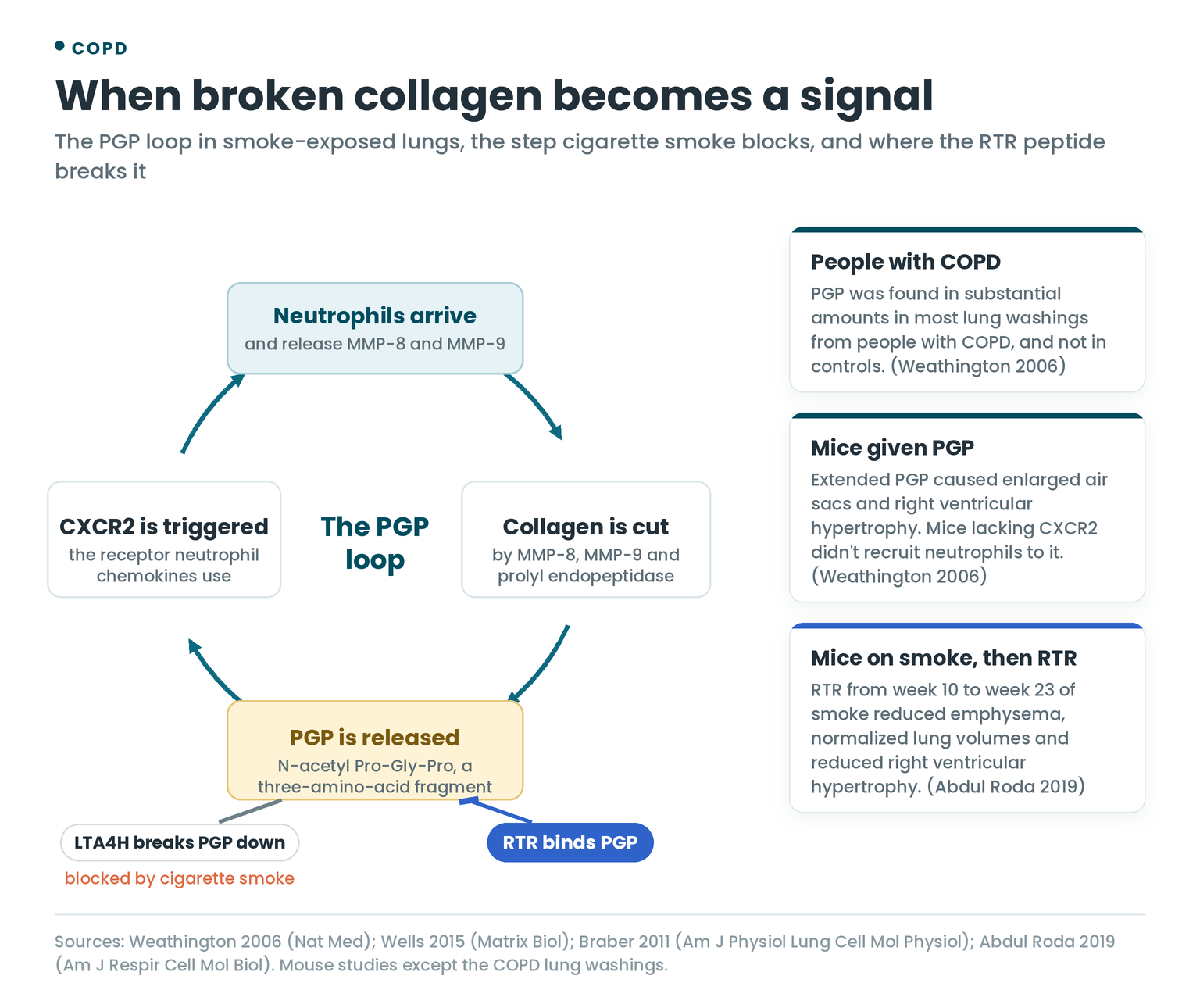
In 2006, Nathaniel Weathington and colleagues described a three-amino-acid fragment of collagen, N-acetyl Pro-Gly-Pro (PGP), that attracts neutrophils. It works through the same CXC receptors as the body’s own neutrophil chemokines: it pulled neutrophils into the lungs of normal mice but not of mice lacking CXCR2. Given for an extended period, it caused alveolar enlargement and right ventricular hypertrophy in mice. PGP was present in substantial concentrations in most lung washings from people with COPD and not in controls.
In a smoker’s lung this becomes a loop. Neutrophils release MMP-8 and MMP-9, which, with prolyl endopeptidase, cut PGP out of collagen, and PGP calls in more neutrophils. The enzyme that normally breaks PGP down, leukotriene A4 hydrolase, is itself inhibited by cigarette smoke. In mice exposed to smoke for 20 weeks, every step of the PGP pathway was elevated, and eight weeks after the smoke stopped they had fallen back or returned to normal.
RTR (arginine-threonine-arginine) is a small peptide that binds PGP. After five days of smoke it reduced the neutrophil influx into mouse lungs. In the longer test from 2019, mice were smoke-exposed for 10 weeks before RTR was started, then kept on smoke and RTR for 13 more weeks. RTR reduced the emphysema, normalized lung volumes and reduced right ventricular hypertrophy. The same paper found that acetylated PGP made human airway cells release more MMP-9 and IL-8.
Elastin Fragments and Macrophages
Elastin gives the alveolar walls their stretch, and its breakdown leaves peptide fragments too, including the six-amino-acid Val-Gly-Val-Ala-Pro-Gly. In 2006, Houghton and colleagues showed that lung washings from smoke-exposed mice attracted monocytes, that the attraction came from elastin fragments, and that it was missing in mice lacking macrophage elastase (MMP-12). An antibody against the fragments blocked smoke-induced monocyte recruitment to the lung, and in an elastase model of emphysema it stopped both the macrophage build-up and the airspace enlargement.
GHK and the Emphysema Gene Signature
Joshua Campbell and colleagues took eight regions from each of eight lungs, six removed at transplant from people with severe COPD and two donor lungs, and measured each region’s emphysema severity on micro-CT: 64 tissue samples in all. They found 127 genes that tracked how damaged a region was. Inflammation genes rose with the damage; tissue-repair genes, including the TGF-beta pathway, fell.
Searching the Connectivity Map, a database of how compounds shift gene expression, for one that reverses that pattern turned up GHK, the tripeptide glycyl-histidyl-lysine. In human lung fibroblasts, GHK switched on TGF-beta-like repair genes and organized the cells’ actin skeleton. In fibroblasts taken from COPD lungs, GHK restored the cells’ ability to contract and remodel collagen. This was cell work on human lung tissue, and the authors called for studies of what the reversal does to disease progression.
Peptides for Lung Fibrosis
Most of the fibrosis work uses bleomycin, a cancer drug whose use is limited by the lung scarring it causes. Given to mice, it produces a fibrosis model in a few weeks.
Caveolin-1 Peptides: From Mice to a Phase 1b Trial
Caveolin-1 is a membrane protein, and a 2006 study found it markedly reduced in lung tissue and lung fibroblasts from people with idiopathic pulmonary fibrosis (IPF). The peptides come from a stretch of it called the scaffolding domain, amino acids 82 to 101, and peptides copying that stretch suppressed bleomycin-induced lung and skin fibrosis in mice. A 2022 study split that stretch into three overlapping eight-amino-acid pieces, and all three were active in bleomycin mice.
A seven-amino-acid version, CSP7, reduced fibrosis that was already established in mice, partly by keeping alveolar epithelial cells alive. LTI-03 is a seven-amino-acid peptide from the same domain, formulated as an inhaled dry powder. In slices of living lung tissue from IPF patients, it reduced collagen staining in a dose-dependent way (a 2024 preprint).
The first trial in patients was published in 2026. Twenty-four people with IPF were randomized to inhaled LTI-03 at one of two doses or placebo for 14 days. There were no severe treatment-emergent adverse events, no treatment-related discontinuations and no airway narrowing on spirometry. In brushings from deep in the airways, both doses lowered interleukin-11 and thymic stromal lymphopoietin compared with placebo, and the higher dose also lowered collagen type 1 alpha 1, the chemokine CXCL7 and galectin-7. Other measured markers didn’t change significantly.
Ac-SDKP and Thymosin Beta-4
Ac-SDKP is a four-amino-acid peptide found throughout the body. Its most likely precursor is thymosin beta-4, from which the enzyme prolyl oligopeptidase cuts it, and it’s broken down almost entirely by ACE.
ACE has two active sites, and in 2010 mice with the N-terminal site switched off resisted bleomycin lung injury. When their Ac-SDKP production was blocked, they scarred like normal mice, and giving Ac-SDKP to normal mice reduced their fibrosis. Because ACE clears it so fast, a 2020 study swapped two of its amino acids for their mirror-image forms. The new version lasted longer in rat and human serum and protected mouse lungs from bleomycin better than the original.
Thymosin beta-4 itself has been tested too. In a 2025 mouse study, nebulized recombinant human thymosin beta-4 reduced bleomycin fibrosis whether it was started early, midway or late, and in cell work it slowed lung fibroblast growth and activation through the TGF-beta1 pathway. TB-500 is a fragment of thymosin beta-4, and I covered how it’s built in Is TB-500 a Steroid?
GHK-Cu in Bleomycin Mice
Bound to copper, GHK becomes GHK-Cu, a form that improves GHK’s bioavailability. In a 2020 study, GHK-Cu injected every other day into bleomycin mice reduced inflammation and collagen deposition at day 21. It lowered TNF-alpha, IL-6 and myeloperoxidase activity in lung washings, rebalanced MMP-9 against its inhibitor TIMP-1, and partly prevented the shift of lung cells toward a scar-forming state through the Nrf2, NF-kB and TGF-beta1/Smad2/3 pathways.
Peptides for ARDS
In acute respiratory distress syndrome, protein-rich fluid leaks into the alveoli, and the lungs’ ability to clear it is often impaired.
Solnatide: A Piece of TNF That Clears Fluid
Solnatide, also called AP301 or the TIP peptide, is a 17-amino-acid cyclic peptide that copies the lectin-like domain at the tip of the TNF molecule. That part of TNF activates ENaC on alveolar cells, the same channel from the airway section, and in the air sacs that’s what pulls fluid out. In rats with high-altitude pulmonary edema, solnatide lowered lung water and the protein leaking into lung washings, and left the barrier between air and blood more intact.
Two small trials have tested it inhaled in ventilated patients:
- Phase IIa, 2017. Forty adults with ARDS got inhaled AP301 or saline twice daily for seven days. The change in extravascular lung water didn’t differ between groups (p = 0.196), and neither did oxygenation, ventilation pressures, lung injury scores or 28-day mortality. In an exploratory post-hoc subgroup with SOFA scores of 11 or higher, lung water and ventilation pressures fell more on AP301.
- COVID-19 ARDS pilot, 2025. Thirty ventilated patients in Vienna were randomized before the trial stopped early for slow recruitment (40 were planned). Median ventilator-free days were 0 in both groups, 11 of 15 on solnatide and 9 of 15 on placebo were alive at day 28, and no adverse event was judged related to treatment.
Thymosin alpha 1 has its own sepsis and COVID-19 trials, which I went through in Thymosin Alpha 1 and Bodybuilding.
VIP: One Peptide Across Three Lung Conditions
Vasoactive intestinal peptide is a neuropeptide that relaxes blood vessels, including the lung’s. As a drug it’s called aviptadil.
Pulmonary Hypertension
In 2003, Petkov and colleagues found VIP deficient in the blood and lung tissue of people with primary pulmonary hypertension, with its receptors increased. In the eight patients they treated, VIP lowered mean pulmonary artery pressure and raised cardiac output and mixed venous oxygen saturation.
A 2008 study gave 20 people with pulmonary hypertension a single inhaled dose during right-heart catheterization. It caused a small, temporary pulmonary vasodilation, without changing systemic blood pressure, and improved stroke volume and mixed venous oxygen saturation. Six of the 20 had their pulmonary vascular resistance fall by more than 20%. Part of the reason the effect is short: enzymes on the lung surface called neutral endopeptidases break VIP down quickly. In isolated rabbit lungs, blocking them made inhaled VIP’s effect bigger and longer.
Sarcoidosis
In sarcoidosis, immune cells in the lungs form granulomas, driven partly by TNF-alpha from lung macrophages. Antje Prasse and colleagues gave 20 patients with active disease nebulized VIP for four weeks in an open phase II study. It was well tolerated, it cut TNF-alpha production by cells washed out of their lungs, and it raised the number of regulatory T cells there. In the lab, VIP turned naive T cells into regulatory ones.
COVID-19 Respiratory Failure
TESICO’s aviptadil analysis covered 461 hospitalized adults with COVID-19 respiratory failure at 28 US sites, given IV aviptadil or placebo as three daily 12-hour infusions. Ninety-four percent were in intensive care. On the trial’s day-90 recovery scale, the odds ratio for a better outcome was 1.11 (95% CI 0.80 to 1.55). By day 90, 38% on aviptadil and 36% on placebo had died, and the safety board stopped the aviptadil comparison for futility.
BPC-157 and the Lungs in Rats
BPC-157’s lung data come from rats. In a 2021 study, rats were given monocrotaline, a compound that injures the lining of the lung’s blood vessels and causes pulmonary hypertension. BPC-157 given from day 1 kept the pulmonary hypertension from developing, and started at day 14, when it was already advanced, it attenuated and then eliminated it. That covered vessel wall thickness, right ventricle pressure and the ECG changes.
In a 2025 study of 24 rats, 45 minutes of blocked blood flow to the hind legs followed by two hours of reperfusion damaged the lungs at a distance. Rats given BPC-157 had less lung edema, less alveolar congestion and higher antioxidant activity in lung tissue. More of BPC-157’s research record is in BPC 157 and Cancer.
The Lung Peptide Research on One Grid
All fifteen findings from this article, laid out by peptide and by lung condition.
The evidence grid
Each filled square is a peptide studied in a lung condition, colored by the strongest kind of evidence behind it. Tap one to see the study.
Swipe the grid sideways to see every condition.
Doses Used in the Research
- LTI-03 (people with IPF): 5 or 10 mg a day by inhalation for 14 days.
- Solnatide (people with COVID-19 ARDS): 100 mg inhaled twice a day for 7 days. The 2017 ARDS trial also dosed twice a day for 7 days.
- Aviptadil IV (TESICO): a 12-hour infusion on each of 3 days, at 600, 1,200 and 1,800 pmol/kg.
- Aviptadil inhaled (pulmonary hypertension): a single 100 µg dose.
- VIP nebulized (sarcoidosis): for 4 weeks.
- GHK-Cu (bleomycin mice): 0.2, 2 or 20 µg per gram of body weight, injected every other day.
- BPC-157 (rats): 10 µg/kg or 10 ng/kg once a day by injection, or continuously in drinking water.
Frequently Asked Questions
What peptides are being studied for lung health?
Your airways make their own antimicrobial peptides, the defensins and cathelicidins. Peptides tested as treatments include the surfactant peptide sinapultide in preterm babies, solnatide and VIP in lung disease patients, LTI-03 in pulmonary fibrosis, and RTR, GHK, Ac-SDKP, thymosin beta-4, GHK-Cu and BPC-157 in animal or cell studies.
Can peptides reverse lung fibrosis?
In mice, CSP7 reduced fibrosis that was already established, and inhaled thymosin beta-4 worked even when started late. In people, LTI-03’s 14-day phase 1b lowered fibrosis-related markers in airway brushings.
Is there a peptide for COPD or emphysema?
The PGP-binding peptide RTR reduced smoke-induced emphysema in mice when started after 10 weeks of smoke. GHK came out of a database search for compounds that reverse the emphysema gene pattern, and it restored collagen remodeling in fibroblasts from COPD lungs.
Does BPC-157 help the lungs?
In rats, it prevented and reversed monocrotaline-induced pulmonary hypertension and reduced lung damage after limb ischemia and reperfusion.
Did VIP (aviptadil) work for COVID-19 respiratory failure?
In the 461-patient TESICO trial, aviptadil didn’t significantly improve day-90 outcomes, and 38% died against 36% on placebo. Earlier small studies found it lowered lung artery pressure in pulmonary hypertension and calmed lung inflammation in sarcoidosis.
What is the KL4 peptide?
It’s sinapultide, a 21-amino-acid chain of repeating leucine and lysine designed to mimic surfactant protein B. As the synthetic surfactant lucinactant, it was tested against other surfactants in two 2005 trials of premature babies.
Stay sharp,
Joe Mars
The Peptide Report
P.S. Reading the lung research for yourself, or following one of these peptides? Bring your questions to the Peptides & Biohacking Hub on Skool and we’ll go through the papers together.
References
- Ochs M, Nyengaard JR, Jung A, et al. The number of alveoli in the human lung. Am J Respir Crit Care Med. 2004;169(1):120-124. doi:10.1164/rccm.200308-1107OC
- Hiemstra PS. The role of epithelial beta-defensins and cathelicidins in host defense of the lung. Exp Lung Res. 2007;33(10):537-542. doi:10.1080/01902140701756687
- Goldman MJ, Anderson GM, Stolzenberg ED, et al. Human beta-defensin-1 is a salt-sensitive antibiotic in lung that is inactivated in cystic fibrosis. Cell. 1997;88(4):553-560. doi:10.1016/s0092-8674(00)81895-4
- Bals R, Wang X, Wu Z, et al. Human beta-defensin 2 is a salt-sensitive peptide antibiotic expressed in human lung. J Clin Invest. 1998;102(5):874-880. doi:10.1172/JCI2410
- Scott DW, Walker MP, Sesma J, et al. SPX-101 is a novel epithelial sodium channel-targeted therapeutic for cystic fibrosis that restores mucus transport. Am J Respir Crit Care Med. 2017;196(6):734-744. doi:10.1164/rccm.201612-2445OC
- Sesma JI, Wu B, Stuhlmiller TJ, Scott DW. SPX-101 is stable in and retains function after exposure to cystic fibrosis sputum. J Cyst Fibros. 2019;18(2):244-250. doi:10.1016/j.jcf.2018.06.002
- Moore PJ, Reidel B, Ghosh A, et al. Cigarette smoke modifies and inactivates SPLUNC1, leading to airway dehydration. FASEB J. 2018;32(12):fj201800345R. doi:10.1096/fj.201800345R
- Moya FR, Gadzinowski J, Bancalari E, et al. A multicenter, randomized, masked, comparison trial of lucinactant, colfosceril palmitate, and beractant for the prevention of respiratory distress syndrome among very preterm infants. Pediatrics. 2005;115(4):1018-1029. doi:10.1542/peds.2004-2183
- Sinha SK, Lacaze-Masmonteil T, Valls i Soler A, et al. A multicenter, randomized, controlled trial of lucinactant versus poractant alfa among very premature infants at high risk for respiratory distress syndrome. Pediatrics. 2005;115(4):1030-1038. doi:10.1542/peds.2004-2231
- Wolfson MR, Wu J, Hubert TL, et al. Lucinactant attenuates pulmonary inflammatory response, preserves lung structure, and improves physiologic outcomes in a preterm lamb model of RDS. Pediatr Res. 2012;72(4):375-383. doi:10.1038/pr.2012.96
- Weathington NM, van Houwelingen AH, Noerager BD, et al. A novel peptide CXCR ligand derived from extracellular matrix degradation during airway inflammation. Nat Med. 2006;12(3):317-323. doi:10.1038/nm1361
- Wells JM, Gaggar A, Blalock JE. MMP generated matrikines. Matrix Biol. 2015;44-46:122-129. doi:10.1016/j.matbio.2015.01.016
- Braber S, Koelink PJ, Henricks PA, et al. Cigarette smoke-induced lung emphysema in mice is associated with prolyl endopeptidase, an enzyme involved in collagen breakdown. Am J Physiol Lung Cell Mol Physiol. 2011;300(2):L255-L265. doi:10.1152/ajplung.00304.2010
- Abdul Roda M, Xu X, Abdalla TH, et al. Proline-glycine-proline peptides are critical in the development of smoke-induced emphysema. Am J Respir Cell Mol Biol. 2019;61(5):560-566. doi:10.1165/rcmb.2018-0216OC
- Houghton AM, Quintero PA, Perkins DL, et al. Elastin fragments drive disease progression in a murine model of emphysema. J Clin Invest. 2006;116(3):753-759. doi:10.1172/JCI25617
- Campbell JD, McDonough JE, Zeskind JE, et al. A gene expression signature of emphysema-related lung destruction and its reversal by the tripeptide GHK. Genome Med. 2012;4(8):67. doi:10.1186/gm367
- Li P, Xiao HD, Xu J, et al. Angiotensin-converting enzyme N-terminal inactivation alleviates bleomycin-induced lung injury. Am J Pathol. 2010;177(3):1113-1121. doi:10.2353/ajpath.2010.081127
- Wang XM, Zhang Y, Kim HP, et al. Caveolin-1: a critical regulator of lung fibrosis in idiopathic pulmonary fibrosis. J Exp Med. 2006;203(13):2895-2906. doi:10.1084/jem.20061536
- Reese CF, Chinnakkannu P, Tourkina E, et al. Multiple subregions within the caveolin-1 scaffolding domain inhibit fibrosis, microvascular leakage, and monocyte migration. PLoS One. 2022;17(2):e0264413. doi:10.1371/journal.pone.0264413
- Venkatesan S, Fan L, Tang H, et al. Caveolin-1 scaffolding domain peptide abrogates autophagy dysregulation in pulmonary fibrosis. Sci Rep. 2022;12(1):11086. doi:10.1038/s41598-022-14832-4
- MacKenzie B, Mahavadi P, Jannini-Sa YAP, et al. Pre-clinical proof-of-concept of anti-fibrotic activity of caveolin-1 scaffolding domain peptide LTI-03 in ex vivo precision cut lung slices from patients with idiopathic pulmonary fibrosis. bioRxiv [preprint]. 2024. doi:10.1101/2024.04.24.589970
- Molyneaux PL, Hirani NA, Chia CCK, et al. Inhaled LTI-03 for idiopathic pulmonary fibrosis: a randomized dose escalation study. Nat Commun. 2026;17(1):7620. doi:10.1038/s41467-026-75291-3
- Cavasin MA, Rhaleb NE, Yang XP, Carretero OA. Prolyl oligopeptidase is involved in release of the antifibrotic peptide Ac-SDKP. Hypertension. 2004;43(5):1140-1145. doi:10.1161/01.HYP.0000126172.01673.84
- Qiu Y, Wang Z, Zhang X, et al. A long-acting isomer of Ac-SDKP attenuates pulmonary fibrosis through SRPK1-mediated PI3K/AKT and Smad2 pathway inhibition. IUBMB Life. 2020;72(12):2611-2626. doi:10.1002/iub.2389
- Yu R, Li S, Chen L, et al. Inhaled exogenous thymosin beta 4 suppresses bleomycin-induced pulmonary fibrosis in mice via TGF-β1 signalling pathway. J Pharm Pharmacol. 2025;77(4):582-592. doi:10.1093/jpp/rgae143
- Mendias CL, Awan TM. Safety and efficacy of approved and unapproved peptide therapies for musculoskeletal injuries and athletic performance. Sports Med. 2026;56(8):1921-1935. doi:10.1007/s40279-026-02437-0
- Ma WH, Li M, Ma HF, et al. Protective effects of GHK-Cu in bleomycin-induced pulmonary fibrosis via anti-oxidative stress and anti-inflammation pathways. Life Sci. 2020;241:117139. doi:10.1016/j.lfs.2019.117139
- Martin-Malpartida P, Arrastia-Casado S, Farrera-Sinfreu J, et al. Conformational ensemble of the TNF-derived peptide solnatide in solution. Comput Struct Biotechnol J. 2022;20:2082-2090. doi:10.1016/j.csbj.2022.04.031
- Zhou Q, Wang D, Liu Y, et al. Solnatide demonstrates profound therapeutic activity in a rat model of pulmonary edema induced by acute hypobaric hypoxia and exercise. Chest. 2017;151(3):658-667. doi:10.1016/j.chest.2016.10.030
- Krenn K, Lucas R, Croizé A, et al. Inhaled AP301 for treatment of pulmonary edema in mechanically ventilated patients with acute respiratory distress syndrome: a phase IIa randomized placebo-controlled trial. Crit Care. 2017;21(1):194. doi:10.1186/s13054-017-1795-x
- Krenn K, Kraft F, Urban M, et al. Efficacy of solnatide to treat pulmonary permeability edema in SARS-CoV-2 positive patients with moderate to severe ARDS: a randomized controlled pilot-trial. Anaesth Crit Care Pain Med. 2025;44(3):101520. doi:10.1016/j.accpm.2025.101520
- Petkov V, Mosgoeller W, Ziesche R, et al. Vasoactive intestinal peptide as a new drug for treatment of primary pulmonary hypertension. J Clin Invest. 2003;111(9):1339-1346. doi:10.1172/JCI17500
- Leuchte HH, Baezner C, Baumgartner RA, et al. Inhalation of vasoactive intestinal peptide in pulmonary hypertension. Eur Respir J. 2008;32(5):1289-1294. doi:10.1183/09031936.00050008
- Leuchte HH, Prechtl C, Callegari J, et al. Augmentation of the effects of vasoactive intestinal peptide aerosol on pulmonary hypertension via coapplication of a neutral endopeptidase 24.11 inhibitor. Am J Physiol Lung Cell Mol Physiol. 2015;308(6):L563-L568. doi:10.1152/ajplung.00317.2014
- Prasse A, Zissel G, Lützen N, et al. Inhaled vasoactive intestinal peptide exerts immunoregulatory effects in sarcoidosis. Am J Respir Crit Care Med. 2010;182(4):540-548. doi:10.1164/rccm.200909-1451OC
- Brown SM, Barkauskas CE, Grund B, et al. Intravenous aviptadil and remdesivir for treatment of COVID-19-associated hypoxaemic respiratory failure in the USA (TESICO): a randomised, placebo-controlled trial. Lancet Respir Med. 2023;11(9):791-803. doi:10.1016/S2213-2600(23)00147-9
- Udovicic M, Sever M, Kavur L, et al. Stable gastric pentadecapeptide BPC 157 therapy for monocrotaline-induced pulmonary hypertension in rats leads to prevention and reversal. Biomedicines. 2021;9(7):822. doi:10.3390/biomedicines9070822
- Demirtaş H, Özer A, Yıldırım AK, et al. Protective effects of BPC 157 on liver, kidney, and lung distant organ damage in rats with experimental lower-extremity ischemia-reperfusion injury. Medicina (Kaunas). 2025;61(2):291. doi:10.3390/medicina61020291