

Somebody shaved both flanks of a rat, rubbed a peptide into one side and plain vehicle into the other, and counted follicles a week later. The treated side had roughly twice as many growing.
That one image is where almost every TB-500 hair claim traces back to. What happened next is stranger than the headline, and it runs through a mouse with odd teeth, a herd of Chinese cashmere goats, and a dish of human scalp.
Where the Hair Claim Started
It started as a side observation in a wound study. Deborah Philp and colleagues at the National Institutes of Health were looking at how thymosin beta-4 affected healing in rat skin, and when they examined the tissue under the microscope they noticed something nobody had gone looking for: the number of hair follicles at the wound margins had gone up after seven days of treatment.
So they ran the obvious follow-up. As Bai Dai and colleagues describe the work in their 2021 review, the sides of healthy rats were shaved, and thymosin beta-4 or control vehicle was applied to each side. One animal, two flanks, its own control. After seven days, the skin treated with the peptide had approximately twice as many growing follicles as the vehicle side.
Two details in that follow-up matter more than the doubling, and they almost never travel with it.
The first is the route. This was applied to the skin surface. Nobody injected anything.
The second is what happened when they stopped. After three weeks of continuous treatment, the new hair persisted for more than thirty days. But within fourteen days of stopping, the follicle count dropped back to its original level. The effect held while the treatment held, and let go after it.
The same group checked whether it worked in mice as well, and eight-week-old C57BL/6 wild-type mice treated with thymosin beta-4 showed rapid hair regeneration, with histology confirming the peptide was pushing follicles through a cycle transition rather than simply making existing hairs longer.
Dai’s review is blunt about the limits of this era of work. The methods, the authors write, were relatively simplistic, and the studies did not fully explain how thymosin beta-4 affects follicle growth and development. That is a fair read of a result that came out of a wound experiment by accident.
What a Follicle Is Doing When It Grows
To see why a peptide could move a follicle at all, it helps to know that a hair follicle is the only organ in the mammalian body that spends your entire life taking itself apart and rebuilding.
Ralf Paus and Kerstin Foitzik laid this out in their tour of what they called the hair cycle clock. The follicle runs from a stage of rapid growth, anagen, into an apoptosis-driven regression called catagen, then through an interspersed period of relative quiescence, telogen, and back into anagen. The clock that drives the cycling, they note, sits inside the follicle itself. Nothing outside it is keeping time.
The rebuilding is done from a reservoir. In 1990, George Cotsarelis, Tung-Tien Sun and Robert Lavker went looking for the follicle’s stem cells and found them somewhere other than where the field expected. Label-retaining cells were not in the matrix down in the hair bulb; they sat exclusively in a region partway up the follicle called the bulge, a subpopulation of outer root sheath cells at the point where the arrector pili muscle attaches. The muscle that raises a goosebump marks the spot. Those keratinocytes are relatively undifferentiated, normally slow cycling, and they mark the lower end of what the authors called the permanent portion of the follicle.
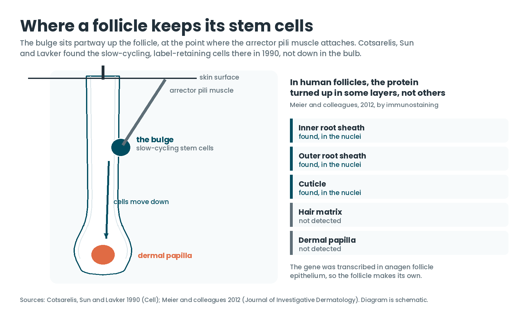
That is the address Philp’s team ended up at. In their 2004 FASEB Journal paper, they reported that a specific subset of follicular keratinocytes in mouse skin expresses thymosin beta-4 in a highly coordinated manner across the hair growth cycle, and that those keratinocytes originate in the bulge. They then isolated clonogenic keratinocytes from rat whisker follicles, cells closely related to the bulge-residing stem cells if not identical to them, and found that nanomolar concentrations of thymosin beta-4 increased both their migration and their differentiation. Expression and secretion of matrix metalloproteinase-2, an enzyme that chews through extracellular matrix, went up as well.
Put together, that is a mechanism with a shape to it: the peptide helps bulge cells and their immediate progeny move down to the base of the follicle, differentiate once they arrive, and clear a path through the surrounding matrix on the way. Our guide to what thymosin beta-4 is and what it does covers the actin biology underneath that, which is the day job this molecule was named for.
The Gene, Not the Vial
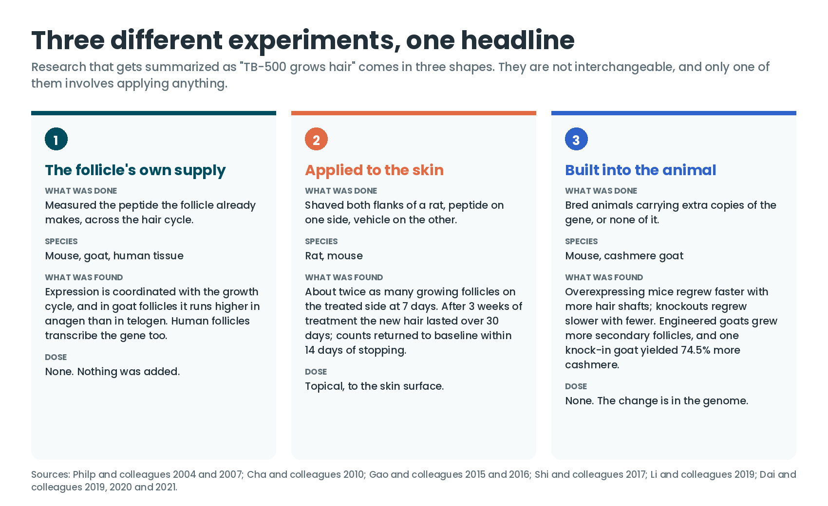
Here is where the literature splits in a way the summaries tend to flatten. Most of the strong, modern results for thymosin beta-4 and hair did not come from applying a peptide to anything. They came from animals built to carry more of the gene, or none of it.
Xiaoyu Gao and colleagues made both. In their 2015 PLOS ONE work they generated mice that overexpressed thymosin beta-4 in the epidermis alongside global knockout mice, plucked the hair, and watched the regrowth. The overexpressing mice regrew faster than controls and the knockouts regrew slower. Histology showed the overexpressing mice carried a higher number of hair shafts, with follicles clustered together into groups where control and knockout follicles stayed separate; shaft numbers in the knockouts were significantly reduced against control. VEGF expression tracked the same direction, up in the overexpressors and down in the knockouts, as did the expression and phosphorylation of p38, ERK and AKT.
The same group went after the upstream switch the following year in Molecular Genetics and Genomics. Using those two mouse lines again, they found beta-catenin and Lef-1, the two key molecules of the Wnt pathway, moving in step with thymosin beta-4. MMP-2 and VEGF rose in the overexpressors and fell dramatically in the knockouts, while E-cadherin stayed put in both. Their reading was that thymosin beta-4 regulates VEGF and MMP-2 through Wnt, beta-catenin and Lef-1, influencing the blood supply around the follicle and activating cell migration. They closed on their own hedge, which is worth carrying intact: thymosin beta-4 may have potential for treating hair growth problems in adults, and its effects should be confirmed in future studies.
Philp’s group had already gone the transgenic route in 2007, reporting hair growth across various rat and mouse models including a thymosin beta-4 overexpressing mouse. Dai’s review fills in that line: the mice were driven off the keratin 5 promoter, and when resting eight-week-old animals were shaved, the overexpressors regrew faster than wild-type mice from the same litter. Hee-Jae Cha and colleagues built the same kind of keratin 5 line in 2010 and found laminin-5 up-regulated in the skin of the transgenic mice. Dai’s review sets that beside earlier work showing laminin-5 is highly expressed in follicles and that follicle integrity depends on integrin beta-1, the major receptor component of laminin-5, so it is a plausible lever rather than an incidental reading.
Cha’s mice are worth sitting with, because the paper’s title names both halves of what happened. Hair growth was accelerated. The transgenic mice also had abnormally shaped white teeth and dull incisors. Keratin 5 is not a hair follicle promoter; it is active across skin epithelium and in developing teeth, and the extra thymosin beta-4 showed up especially around hair follicles and in the teeth. Turn a gene up across a whole tissue compartment and you get everything that compartment does.
The Cashmere Program
If you want to find where thymosin beta-4 and hair have been pushed hardest, it is not a hair loss clinic. It is animal husbandry.
Cashmere is the undercoat a goat grows from its secondary hair follicles, and a goat’s yield is largely a question of how many secondary follicles it has. That makes any molecule that raises secondary follicle counts commercially interesting, and two separate Chinese groups have spent years engineering thymosin beta-4 into goats to find out.
Bingbo Shi and colleagues took the transposon route in 2017, putting the cashmere goat thymosin beta-4 coding sequence behind a hair-follicle-specific promoter, keratin-associated protein 6.1, and generating goats by somatic cell nuclear transfer. Skin histology showed the transgenic goats carried a higher secondary-to-primary follicle ratio than wild type, which is the structural change you would want if the goal is more undercoat per animal.
Xiaocong Li and colleagues went further in 2019, using CRISPR/Cas9 to knock the thymosin beta-4 gene into the goat CCR5 locus without screening or fluorescent markers. The number from that paper is the one that travels: an increase in cashmere yield of 74.5%, with fineness and quality unaffected. It is worth knowing how that number is reported, though, because the paper describes the result in the singular. This was the goat, one knock-in animal, examined closely rather than a herd compared against a herd. RNA sequencing pointed at vasoconstriction, angiogenesis and vascular permeability around the secondary follicles as the processes involved.
The longer-running effort is Bai Dai’s. Writing in Animals at the end of 2019, that group reported having spent the past ten years evaluating thymosin beta-4 by establishing a flock of fifteen cashmere goats engineered to overexpress the gene specifically in their hair follicles. Those goats had more secondary follicles than wild type and produced more cashmere. Pairing the skin transcriptome with the proteome, the authors proposed that thymosin beta-4 works through an interaction with keratin type II cytoskeletal 4 epidermal to mediate ERK signaling.
The same lab also came at it from the other end, screening for genes that differ between a goat follicle in anagen and the same follicle in telogen. Thymosin beta-4 fell out of that screen in the 2020 International Journal of Molecular Sciences paper for a simple reason: its expression in anagen was higher than in telogen. When they then overexpressed it in secondary follicle dermal papilla cells, proliferation went up; when they silenced it, proliferation went down.
What each experiment actually switched on
The hair results for thymosin beta-4 come from very different setups. Pick one to see what drove the gene, where it acted, and what the researchers observed.
Schematic. Shading marks tissue the source describes, not a measured map.
Teaching aid only. Every entry is drawn from the published description of that experiment.
The One Human Experiment
Human hair research has an assay problem, and the field solved it with organ culture. Jennifer Kloepper and colleagues, writing in Experimental Dermatology, call the organ culture of human scalp follicles the best currently available assay for hair research in the human system. A follicle is microdissected out of scalp skin and kept alive in serum-free medium, where you can measure how fast the shaft elongates and whether the follicle holds anagen or tips into catagen. Because anagen VI and early catagen look deceptively similar down a microscope, that paper built the classification system the field now uses: seven qualitative criteria drawn from the morphology of the hair matrix, the dermal papilla and the distribution of pigmentary markers, plus ten quantitative parameters, validated against a known topical hair growth inhibitor to confirm it caught premature catagen when premature catagen was there.
In 2012, Natalia Meier, Dorothee Langan, Ralf Paus and colleagues put thymic peptides into that assay.
They checked expression first. Reverse transcription PCR on the epithelial part of female anagen follicles returned strong, specific signals for thymosin beta-4, so human follicles make it themselves. By immunostaining, the protein turned up only in the nuclei of the inner root sheath, outer root sheath and cuticle, not in the matrix or the dermal papilla.
Then they added it. Follicles were treated for seven days with thymosin beta-4 at 100 or 1,000 ng/ml, and hair shaft production came in 10 to 20% lower than vehicle-treated controls. At 1,000 ng/ml, the peptide shortened the duration of anagen and prematurely induced catagen. Proliferation in the hair matrix was not the route; counting Ki67-positive cells across anagen follicles showed no change against control.
The peptide that did stimulate growth in the same experiment was a different one. Thymulin at 10 pg/ml increased the shaft growth rate, kept follicles in anagen VI longer, raised Ki67-positive cells in the matrix and reduced the number of apoptotic cells. That anagen-prolonging effect missed significance at seven days, where the comparison returned a P-value of 0.06, and reached significance in nine-day cultures. The authors named thymulin, not thymosin beta-4, as their clinically interesting candidate hair growth stimulator, and framed the overall finding as selected thymic peptides being exploitable for stimulating and also for inhibiting human hair growth.
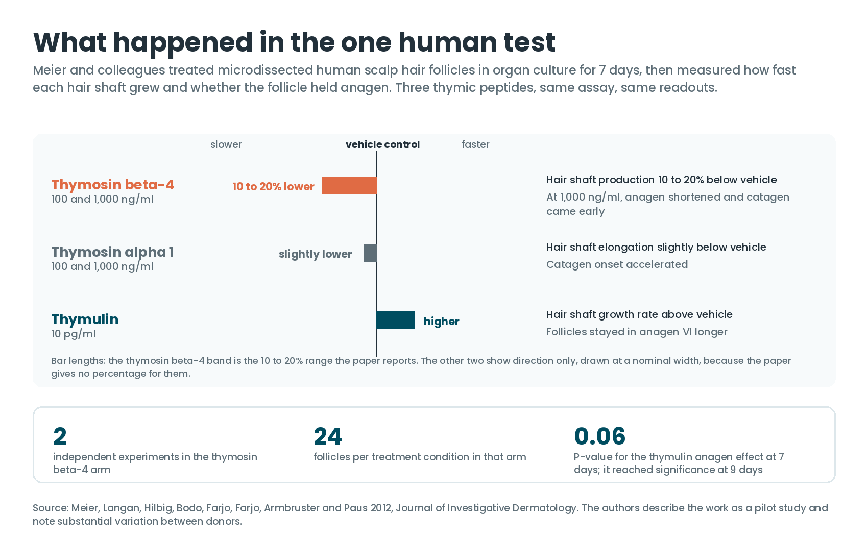
Hold that result at the size it is. The thymosin beta-4 arm ran across two independent experiments with twenty-four follicles per treatment condition, against three experiments and thirty-six follicles for the 10 pg/ml thymulin and the thymosin alpha 1 arms. The authors call it a pilot study, and they flag substantial variation between donors in how follicles responded. Seven days in a dish is also not a scalp. A microdissected follicle sits in serum-free medium rather than in a blood supply, which matters when the mechanism proposed in rodents runs partly through angiogenesis around the follicle.
The assay registers movement in both directions. The same laboratory, in the same organ culture system, has used these readouts to identify other molecules that shorten anagen, including the neuropeptide galanin at 100 nmol/L over nine days and dopamine at 10 to 1,000 nmol/L over seven.
Which Way a Follicle Points
One more finding deserves its own paragraph, because it is about a property of hair nobody thinks to measure: direction.
Krishnanand Padmanabhan and colleagues, working in mouse embryos, depleted thymosin beta-4 and found that follicle angling went wrong. Hair follicles do not grow straight out of skin; they lie at an angle, and that angle is set by planar cell polarity, the system that gives a sheet of cells a shared sense of which way is which. Depleting the peptide hindered eyelid closure and disrupted follicle angling through polarity defects. The cause traced back to adherens junctions, whose structural organization and stability fell apart because of changes in how globular and filamentous actin were distributed. In cultured keratinocytes, the depletion raised the ratio of globular to filamentous actin near the junctions and reduced how much globular actin got built into junctional actin networks, without changing overall actin expression or total filamentous actin. A drug that shifted that same ratio reproduced the effect.
That is the actin-handling job the molecule is named for, showing up in skin as the geometry of a hair.
Doses Used in the Research
Doses only mean something attached to their species and their route, so they are listed that way. For what the numbers on a vial label actually translate to, our piece on blend ratios and vial math covers the arithmetic.
- Rat skin, topical: thymosin beta-4 or vehicle applied to shaved flanks, seven days to the follicle-count read-out, with a three-week continuous-treatment arm (Philp 2004, as described by Dai 2021).
- Rat whisker follicle keratinocytes, in culture: nanomolar concentrations increased migration and differentiation (Philp 2004).
- Mice, topical: eight-week-old C57BL/6 wild-type animals (Dai 2021).
- Human scalp follicles, organ culture: 100 and 1,000 ng/ml thymosin beta-4 for seven days (Meier 2012).
- Human scalp follicles, same experiment, other peptides: thymulin at 10 and 100 pg/ml; thymosin alpha 1 at 100 and 1,000 ng/ml (Meier 2012).
- Transgenic and knockout animals: no dose. Mouse lines were driven by the keratin 5 promoter (Philp 2007, Cha 2010); goats by the keratin-associated protein 6.1 promoter (Shi 2017) or a knock-in at the CCR5 locus (Li 2019).
Every route in this set was topical, in culture, or genetic.
Frequently Asked Questions
Is TB-500 the same thing as thymosin beta-4?
Not quite, and the distinction matters when you are reading studies. Every paper described here tested thymosin beta-4 itself, the 43-amino-acid protein. Our piece on how TB-500 compares with steroids walks through what the research-chemical name covers.
Was the hair research done with injections?
No. The rodent work applied thymosin beta-4 to the skin surface, and the human work added it to follicles in a dish. The transgenic animals carried extra copies of the gene instead of receiving a peptide.
How long did the rodent effect last?
After three weeks of continuous topical treatment, the new hair persisted for more than thirty days. Follicle counts returned to their starting level within fourteen days of stopping treatment.
What happened when thymosin beta-4 was put on human hair follicles?
In the Meier 2012 organ culture study, hair shaft production ran 10 to 20% below vehicle controls, and at 1,000 ng/ml the peptide shortened anagen and brought on catagen early. Thymulin, tested alongside it, increased shaft growth and prolonged anagen.
Why do the goat studies report such large numbers?
Because they measure a different thing. Cashmere yield depends on secondary hair follicle density, and the goats were engineered to express thymosin beta-4 in their follicles from birth rather than treated with a peptide. The 74.5% yield increase comes from a single CRISPR knock-in animal.
Does thymosin beta-4 affect anything about hair besides growth rate?
Mouse embryo work found it is needed for follicles to be angled correctly, through its role in planar cell polarity and adherens junction stability.
References
- Philp D, Nguyen M, Scheremeta B, St-Surin S, Villa AM, Orgel A, Kleinman HK, Elkin M. Thymosin beta4 increases hair growth by activation of hair follicle stem cells. FASEB J. 2004;18(2):385-387. doi:10.1096/fj.03-0244fje
- Philp D, Goldstein AL, Kleinman HK. Thymosin beta4 promotes angiogenesis, wound healing, and hair follicle development. Mech Ageing Dev. 2004;125(2):113-115. doi:10.1016/j.mad.2003.11.005
- Philp D, St-Surin S, Cha HJ, Moon HS, Kleinman HK, Elkin M. Thymosin beta 4 induces hair growth via stem cell migration and differentiation. Ann N Y Acad Sci. 2007;1112:95-103. doi:10.1196/annals.1415.009
- Cha HJ, Philp D, Lee SH, Moon HS, Kleinman HK, Nakamura T. Over-expression of thymosin beta 4 promotes abnormal tooth development and stimulation of hair growth. Int J Dev Biol. 2010;54(1):135-140. doi:10.1387/ijdb.082735hc
- Meier N, Langan D, Hilbig H, Bodó E, Farjo NP, Farjo B, Armbruster FP, Paus R. Thymic peptides differentially modulate human hair follicle growth. J Invest Dermatol. 2012;132(5):1516-1519. doi:10.1038/jid.2012.2
- Gao X, Liang H, Hou F, Zhang Z, Nuo M, Guo X, Liu D. Thymosin beta-4 induces mouse hair growth. PLoS One. 2015;10(6):e0130040. doi:10.1371/journal.pone.0130040
- Gao XY, Hou F, Zhang ZP, Nuo MT, Liang H, Cang M, Wang ZG, Wang X, Xu T, Yan LY, Guo XD, Liu DJ. Role of thymosin beta 4 in hair growth. Mol Genet Genomics. 2016;291(4):1639-1646. doi:10.1007/s00438-016-1207-y
- Shi B, Ding Q, He X, Zhu H, Niu Y, Cai B, Cai J, Lei A, Kang D, Yan H, Ma B. Tβ4-overexpression based on the piggyBac transposon system in cashmere goats alters hair fiber characteristics. Transgenic Res. 2017;26(1):77-85. doi:10.1007/s11248-016-9988-7
- Li X, Hao F, Hu X, Wang H, Dai B, Wang X, Liang H, Cang M, Liu D. Generation of Tβ4 knock-in cashmere goat using CRISPR/Cas9. Int J Biol Sci. 2019;15(8):1743-1754. doi:10.7150/ijbs.34820
- Dai B, Liang H, Guo DD, Bi ZW, Yuan JL, Jin Y, Huan L, Guo XD, Cang M, Liu DJ. The overexpression of Tβ4 in the hair follicle tissue of Alpas cashmere goats increases cashmere yield and promotes hair follicle development. Animals (Basel). 2019;10(1):75. doi:10.3390/ani10010075
- Dai B, Hao F, Xu T, Zhu B, Ren LQ, Han XY, Liu DJ. Thymosin β4 identified by transcriptomic analysis from HF anagen to telogen promotes proliferation of SHF-DPCs in Albas cashmere goat. Int J Mol Sci. 2020;21(7):2268. doi:10.3390/ijms21072268
- Dai B, Sha RN, Yuan JL, Liu DJ. Multiple potential roles of thymosin β4 in the growth and development of hair follicles. J Cell Mol Med. 2021;25(3):1350-1358. doi:10.1111/jcmm.16241
- Padmanabhan K, Grobe H, Cohen J, Soffer A, Mahly A, Adir O, Zaidel-Bar R, Luxenburg C. Thymosin β4 is essential for adherens junction stability and epidermal planar cell polarity. Development. 2020;147(23):dev193425. doi:10.1242/dev.193425
- Paus R, Foitzik K. In search of the “hair cycle clock”: a guided tour. Differentiation. 2004;72(9-10):489-511. doi:10.1111/j.1432-0436.2004.07209004.x
- Cotsarelis G, Sun TT, Lavker RM. Label-retaining cells reside in the bulge area of pilosebaceous unit: implications for follicular stem cells, hair cycle, and skin carcinogenesis. Cell. 1990;61(7):1329-1337. doi:10.1016/0092-8674(90)90696-c
- Kloepper JE, Sugawara K, Al-Nuaimi Y, Gáspár E, van Beek N, Paus R. Methods in hair research: how to objectively distinguish between anagen and catagen in human hair follicle organ culture. Exp Dermatol. 2010;19(3):305-312. doi:10.1111/j.1600-0625.2009.00939.x
- Holub BS, Kloepper JE, Tóth BI, Bíró T, Kofler B, Paus R. The neuropeptide galanin is a novel inhibitor of human hair growth. Br J Dermatol. 2012;167(1):10-16. doi:10.1111/j.1365-2133.2012.10890.x
- Langan EA, Lisztes E, Bíró T, Funk W, Kloepper JE, Griffiths CEM, Paus R. Dopamine is a novel, direct inducer of catagen in human scalp hair follicles in vitro. Br J Dermatol. 2013;168(3):520-525. doi:10.1111/bjd.12113
Stay sharp,
Joe Mars
The Peptide Report
P.S. The sharper questions on this one — why a seven-day dish result and a three-week rodent result are not the same claim, and what either would need to become one — usually get worked out with other people. We talk about it over at the Peptides & Biohacking Hub.