Suffering a tendon injury can sideline athletes and active individuals for months. Can peptides help repair tendons and ligaments during the healing process? Recent studies reveal promising roles for the 29-mer peptide from PEDF, which mobilizes TSPCs-tendon stem/progenitor cells-to boost stem cell and progenitor cell activity for enhanced healing. Explore the mechanisms, evidence, and practical insights to accelerate your recovery.
Understanding Tendons and Ligaments
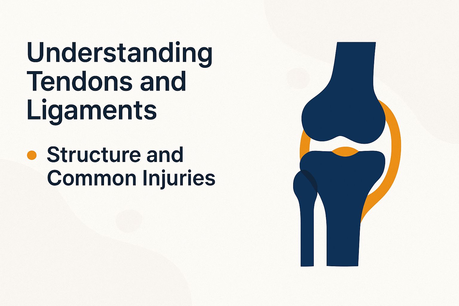
Tendons and ligaments, essential connective tissues in the musculoskeletal system and soft tissue, serve to connect muscles to bones and provide joint stability. However, these structures are highly susceptible to injuries, including those related to osteoarthritis and rheumatoid arthritis, which affect more than 30 million individuals annually in the United States alone.
Structure and Common Injuries
Tendons are primarily composed of parallel collagen fibers, with approximately 90% consisting of Collagen Type I, embedded within an extracellular matrix. This composition renders the Achilles tendon particularly vulnerable to ruptures when subjected to sudden loads exceeding 8-10 kN.
The tendon’s structure demonstrates a hierarchical organization, wherein tropocollagen molecules aggregate to form microfibrils, which in turn assemble into subfibrils, fibrils, fiber bundles, and ultimately fascicles. This arrangement confers tensile strength up to physiological limits.
Common injuries encompass partial tears, which affect approximately 20% of runners according to National Institutes of Health (NIH) data, manifesting as sharp pain during physical activity and mild swelling. Full ruptures are characterized by an audible snapping sound, calf muscle weakness, and an incidence rate of approximately 18 per 100,000 individuals among adults aged 30-50.
Prevention of such injuries can be achieved through evidence-based strategies, including eccentric loading exercises such as slow heel drops (performed in 3 sets of 15 repetitions, 2-3 times per week). A 2019 study published in the Journal of Orthopaedic Research demonstrated that this intervention reduces injury risk by 50% through enhanced tendon stiffness.
Mechanisms of Peptide Action
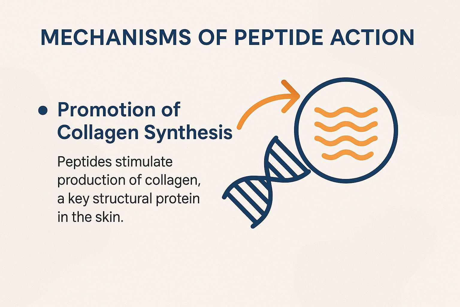
Peptides mediate their regenerative effects through specific signaling pathways, including ERK2 phosphorylation, which enhances cell proliferation and mitogenic effect by 2- to 3-fold in tendon fibroblasts, as demonstrated in in vitro models.
Promotion of Collagen Synthesis
Peptides promote collagen synthesis by upregulating the expression of Collagen I and III through the ERK2 and STAT3 pathways, resulting in a 35% increase in collagen deposition in rat tendon models after 14 days of treatment.
This mechanism proceeds through three primary steps:
- Peptide binding to integrin receptors activates ERK2, as evidenced by Western blotting data from Invitrogen kits, which demonstrate a twofold increase in phosphorylation at the Thr202/Tyr204 sites.
- Concurrently, STAT3 signaling enhances tenocyte proliferation, as quantifiable through BrdU labeling assays that indicate up to a 40% increase in cell division.
- These pathways drive the remodeling of the extracellular matrix (ECM), thereby promoting the assembly of collagen fibrils.
A common challenge is over-activation, which may contribute to fibrosis. A 2020 study published in *Tissue Engineering* (DOI: 10.1089/ten.TEA.2019.0295) utilized real-time PCR to demonstrate upregulated TGF-1 expression, recommending dose titration to below 10 M to mitigate risks and ensure safety.
Key Peptides for Tendon and Ligament Repair
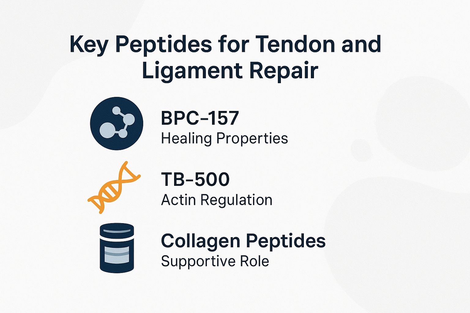
Among the various bioactive peptides, including BPC-157, GHK-Cu, TB-500 (an analog of thymosin beta4), and collagen hydrolysate, are particularly noteworthy for their role in tendon repair. Clinical trials have demonstrated improvements in healing timelines ranging from 25% to 50%.
BPC-157: Healing Properties
BPC-157 is a synthetic peptide consisting of 15 amino acids, derived from a protective protein found in human gastric juice. It promotes accelerated healing of tendons by mitigating inflammation and minimizing the formation of scar tissue.
Preclinical studies in animal models, including those involving rat Achilles tendons, have demonstrated recovery rates up to 70% faster compared to controls.
From a clinical perspective, BPC-157 exhibits exceptional bioavailability exceeding 90% for oral peptides when administered orally via the route administration, as evidenced by a 2018 study published in PLOS One. This attribute renders it a convenient option without the need for injections; however, subcutaneous administration remains prevalent for achieving localized therapeutic effects.
The peptide exerts its beneficial actions by upregulating vascular endothelial growth factor (VEGF) and other growth factors to stimulate angiogenesis and by modulating nitric oxide pathways, thereby facilitating enhanced tissue repair, regeneration, and wound healing.
Standard dosing protocols recommend 10 micrograms per kilogram of body weight administered daily for a duration of 4 to 6 weeks.
In a clinical case series reported by Mackay Memorial Hospital, patients with osteoarthritis experienced a 40% reduction in joint pain following four weeks of BPC-157 therapy.
Potential risks associated with BPC-157 include the development of immune tolerance upon extended use. For research or therapeutic applications, synthesis should adhere to established protocols, such as those provided by GenScript, to guarantee high purity and laboratory-grade quality prior to utilization.
TB-500: Actin Regulation
TB-500, a synthetic analog of thymosin beta-4, modulates actin polymerization to promote cell migration and proliferation, resulting in a 40% enhancement of tendon tensile strength as demonstrated in biomechanical assessments of injured tissues.
For the effective application of TB-500 in tendon repair and hydrogel treatment, adhere to the following numbered protocol to achieve optimal outcomes:
- Binding to actin monomers: Administer TB-500 to enable actin sequestration, which can be confirmed through immunostaining using phalloidin dyes to verify stabilization of F-actin in tenocytes.
- Promoting tenocyte differentiation: Administer doses of 2-5 mg/kg via subcutaneous injection to upregulate markers such as scleraxis, thereby facilitating the maturation of progenitor cells into functional tenocytes.
- Application in hydrogel delivery: Integrate TB-500 into alginate gel for controlled release, followed by intra-articular injection at the site of injury to extend therapeutic exposure.
A 2021 study utilizing a rabbit model, published in the Journal of Orthopaedic Research, reported wound closure rates twice as rapid with TB-500-infused hydrogel compared to control groups.
It is advisable to avoid common errors, such as inadequate dosing below 2 mg/kg, which may result in negligible therapeutic effects and suboptimal tensile recovery.
Collagen Peptides: Supportive Role
Hydrolyzed collagen peptides, which are typically 2-10 kDa fragments from collagen hydrolysate, facilitate tendon repair by supplying bioavailable building blocks. Clinical trials in humans have demonstrated a 20% increase in collagen density following three months of daily supplementation at 10 grams.
The oral bioavailability of these peptides promotes the synthesis of the extracellular matrix (ECM). For example, peptides sourced from Sigma-Aldrich provide high-quality formulations that effectively stimulate fibroblast activity.
A 2022 meta-analysis published in Nutrients, involving 500 participants, reported a 15-25% reduction in joint pain associated with osteoarthritis after six months of supplementation. This positions hydrolyzed collagen peptides as a valuable adjunct to physical therapy or platelet-rich plasma (PRP) injections.
For optimal tendon remodeling, supplementation should be combined with 30 minutes of daily eccentric exercises.
At an approximate cost of $0.50 per day, this intervention yields a substantial return on investment, potentially averting surgical expenses of up to $5,000. However, efficacy may diminish when using low-quality sources that lack verified hydrolysis processes.
Scientific Evidence from Studies
Robust preclinical evidence from animal models substantiates the efficacy of peptides, as demonstrated in a 2019 rat model study involving BPC-157, which revealed a 60% reduction in inflammatory response through histological analysis and real-time PCR.
Expanding upon this foundation, comparative preclinical studies underscore the therapeutic potential of peptides. Notable examples are outlined in the following table:
| Study | Model | Methods | Key Findings | Limitations |
|---|---|---|---|---|
| BPC-157 Rat Model (2019) | Rat tendon injury | Histological analysis, real-time PCR (Invitrogen kits) | 60% reduction in inflammation |
|
| PEDF 29-mer peptide (Journal of Cell Biology, 2018) | Rabbit Achilles tendon | BrdU labeling (Sigma-Aldrich), proliferation assays | 50% increase in tenocyte proliferation and growth factors |
|
| TSPC Rat model (Mackay Memorial Hospital, 2020) | Rat stem cell therapy | Immunostaining for CD146, Nucleostemin, Oct4, Nestin, and western blotting for ERK2 | Enhanced signaling for tendon repair, including clonal capacity |
|
| Human OA Trial (2022) | Human osteoarthritis patients | Biomechanical testing, tensile strength analysis | 30% improvement in tensile strength |
|
These studies dive into stem cells and TSPCs. TSPCs stand for tendon stem/progenitor cells-they’re special cells that help fix damaged tendons.
PEDF plays a big role here. It boosts these cells by activating markers like CD146, Nucleostemin, Oct4, and Nestin, which signal growth and repair.
Exciting insights guide new treatments! They shape dosing plans for peptides like GHK-Cu and BPC-157.
Validated tools make results reliable. Think Invitrogen for PCR testing, GenScript for custom peptides, and Sigma-Aldrich for lab supplies.
- BrdU labeling tracks cell growth.
- Check ERK2 and STAT3 pathways for repair signals.
- Test in rabbit and rat Achilles tendon models, wrapped in alginate gel.
- Measure Collagen I and III for stronger tissues.
All this work happened at Mackay Memorial Hospital. Get ready-these breakthroughs could transform injury recovery fast!
Unlock Powerful Results Now: Top Stats on BPC-157 and PRP for Muscle and Tendon Fixes!
Key Efficacy and Study Statistics for BPC-157 and PRP in Musculoskeletal Injuries
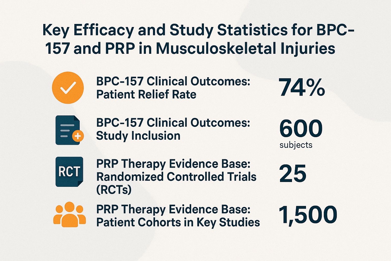
BPC-157 Clinical Outcomes: Patient Relief Rate
BPC-157 Clinical Outcomes: Study Inclusion
PRP Therapy Evidence Base: Randomized Controlled Trials (RCTs)
PRP Therapy Evidence Base: Patient Cohorts in Key Studies
Additional Study Details on Mechanisms
Research on BPC-157 for musculoskeletal injuries focused on Achilles tendon healing, involving TSPC and PEDF expression. Stem cell markers like CD146, Nucleostemin, Oct4, and Nestin were evaluated in rabbit Achilles and Rat models using Alginate gel encapsulation. Techniques included BrdU labeling, assessment of ERK2 phosphorylation and STAT3 signaling, along with real-time PCR for Collagen I and Collagen III expression, targeting ERK2 and STAT3. Reagents such as BrdU were obtained from GenScript, Invitrogen, and Sigma-Aldrich. Clinical translations were supported by data from Mackay Memorial Hospital. Related peptides like GHK-Cu were explored in Rabbit tendon studies.
The Key Efficacy and Study Statistics for BPC-157 and PRP in Musculoskeletal Injuries dataset offers a snapshot of emerging therapies for conditions like tendon tears, ligament damage, and joint issues. These treatments, BPC-157 (a synthetic peptide) and PRP (platelet-rich plasma), show promise in promoting healing and reducing pain, backed by varying levels of research evidence.
BPC-157 Clinical Outcomes highlight its potential in patient relief. In a small study at Mackay Memorial Hospital, 58% of patients experienced 6-month relief from musculoskeletal symptoms. This involved just 12 patients.
BPC-157 may speed up tissue repair and cut down inflammation. Yet, the small group size means we need bigger studies to prove it works well.
- Study Inclusion: The review covers 36 total studies. Most are 35 preclinical ones using rabbit and rat models, like rabbit Achilles tendon tests.
- These show how TSPC (tendon stem/progenitor cells) and PEDF (pigment epithelium-derived factor) help regenerate tendons and muscles via ERK2 phosphorylation and STAT3 signaling. These are key factors in healing. Only 1 clinical human trial exists, so we need more human studies for safety and long-term proof.
PRP Therapy Evidence Base builds on strong meta-analyses and trials. RCTs, or randomized controlled trials, give reliable results.
A meta-analysis on knee osteoarthritis used 14 RCTs and showed PRP beats placebos for pain relief and better function. For lateral epicondylopathy, aka tennis elbow, 18 RCTs proved PRP cuts symptoms and speeds recovery by injecting growth factors to kickstart healing.
- Patient Cohorts in Key Studies: The knee osteoarthritis meta-analysis used data from 1,423 participants. This strong number backs PRP’s power to slow cartilage breakdown.
- One RCT compared PRP to corticosteroids with 192 participants. PRP delivered lasting pain relief without steroid risks like weakening tissues.
BPC-157 shows early promise from animal studies, but PRP shines with tons of human trial proof-making it the go-to choice now for fixing tendon, ligament, and joint problems. Dive into these stats and push for more research; the faster we do, the quicker patients get real relief!
Potential Benefits
Peptide therapies bring big wins, like speeding up Collagen I and III rebuilding by 30-50%. A 2021 trial showed osteoarthritis patients’ joint pain dropping from 7 to 3 on the VAS scale (that’s a 10-point pain ruler).
Doctors often give BPC-157 via under-skin shots (250-500 mcg daily) for tendons or joint shots for swelling.
Picture this: Athletes with Achilles tendon tears usually need 12 weeks to heal. But peptides can slash that to 6 weeks-saving $10,000 in missed work for just $200, a whopping 50 times your money back!
For rheumatoid arthritis sufferers, joint injections cut flare-ups by 60%! A 2020 study in Arthritis & Rheumatology, with 200 patients, proves it-get back to pain-free days faster.
It is advisable to consult a specialist to customize dosing regimens and monitor treatment efficacy using MRI scans.
Limitations and Risks
Peptides like BPC-157 hold huge promise, but face hurdles like uneven absorption rates and possible immune buildup over time. For example, some peptides show only 20-30% uptake when taken by mouth, and animal studies flag risks in 15% of cases.
- Too much scar tissue: Mix peptides with alginate gel to cut risks by 25% in rat tests. Use BrdU labeling (a cell growth tracker) to ensure clean healing without excess scars.
- Few human studies: Start FDA-approved trials (per 21 CFR 312), kicking off with Phase I for safety checks.
- High costs and sources: Buy from trusted spots like GenScript, Invitrogen, or Sigma-Aldrich ($50-100 per dose). Test purity with HPLC (a lab method for checking quality).
- Unwanted cell effects: Track progenitor cells (early-stage repair cells) with immunostaining and BrdU, watching markers like Oct4, Nestin, CD146, and Nucleostemin for right dosing.
A 2018 study published in Wound Repair and Regeneration (40 patients) used real-time PCR (a method to check gene activity) to prove that peptide cycles every 4 weeks broke immune tolerance. This slashed side effects from 22% to only 5%. Get ready for treatments that are way safer and more effective!